

 +44-74-1148-3554




Research Article - Archives of Clinical and Experimental Surgery (2026)
Femoral Supracondylar Dome Osteotomy for Correction of Genu Valgum Deformity: Comparative Analysis of K-Wire Versus Plate Osteosynthesis
Anshul Meena*, Manish Bajaj and Gaurav JainAnshul Meena, Department of Orthopaedic Surgery, LNCT Medical College, Indore, India, Email: dranshulm170@gmail.com
Received: 15-Jun-2026, Manuscript No. EJMACES-26-192733; Editor assigned: 17-Jun-2026, Pre QC No. EJMACES-26-192733; Reviewed: 01-Jul-2026, QC No. EJMACES-26-192733; Revised: 10-Jul-2026, Manuscript No. EJMACES-26-192733; Published: 17-Jul-2026
Abstract
Background: Genu valgum deformity significantly impacts lower limb biomechanics and function. Dome osteotomy represents an effective corrective technique, but the optimal fixation method remains debated. This study compares outcomes of K-wire versus plate osteosynthesis for dome osteotomy fixation.
Methods: Prospective cohort study of 18 patients (22 knees) with symptomatic genu valgum undergoing femoral supracondylar dome osteotomy. Fixation was with either 4 K-wires (2 medial, 2 lateral; n=10 knees) or 3.5 mm locking compression plate (n=12 knees). K-wires were preferentially used in patients with comorbidities (renal rickets, hypophosphatemic rickets) to reduce operative time and blood loss. Preoperative and postoperative assessments included full-length scanograms, radiological analysis, and Bostman functional scores at 6 months follow-up.
Results: Both groups achieved excellent deformity correction with mean tibiofemoral angle reducing from 21.2° to 4.1° (p<0.0001). Union was achieved in all cases, though delayed union occurred in 3 knees (13.6%), all in the K-wire group. K-wire patients required 6-week immobilization versus 3-week immobilization in the plate group before active range of motion. Weight-bearing was initiated at 6-8 weeks in both groups. Complications included peroneal nerve paresis in 1 patient (recovered by 8 weeks), pin tract infections in 4 knees (4/10 K-wire cases, 40%), and superficial infection in 1 plate patient. Bostman scores were excellent or good in 100% of cases at 6 months. Scanogram measurements confirmed no limb length discrepancy in either group.
Conclusions: Dome osteotomy with either K-wire or plate fixation provides reliable correction of genu valgum with excellent functional outcomes. Plate fixation offers earlier active range of motion and reduced infection risk, making it preferable in uncomplicated cases. K-wire fixation remains a viable option for patients with comorbidities requiring operative time reduction, though pin tract infections necessitate rigorous wound care protocols.
Introduction
Genu valgum, commonly referred to as knock-knee deformity, represents a significant coronal plane malalignment of the lower limb. The tibiofemoral angle exceeding 12-15 degrees is considered pathological. The prevalence of significant genu valgum varies geographically, with higher incidence in developing countries where metabolic bone disease, particularly nutritional rickets and hypophosphatemic rickets, remains endemic [1-3].
The etiology of genu valgum is heterogeneous, encompassing metabolic bone disease, post- traumatic sequelae, skeletal dysplasias, and idiopathic presentation. Anatomical localization of deformity is crucial for appropriate surgical correction. Genu valgum typically originates from the distal femur in approximately 60-70% of cases.
Uncorrected genu valgum leads to significant biomechanical abnormalities including abnormal gait, functional disturbances, premature knee osteoarthritis, patellar maltracking, and anterior knee pain. In skeletally mature adolescents and young adults with fixed deformities, corrective osteotomy remains the definitive treatment.
Dome osteotomy is a cylindrical osteotomy wherein bone cuts rotate around a central axis. When the Center of Rotation of Angulation (CORA) is placed at the knee joint level, focal dome osteotomy allows pure correction without translational component or limb length alteration. The technique provides large bony contact surfaces favoring healing [4-6].
While the dome osteotomy technique is wellestablished, the optimal method of fixation remains debated. Internal fixation options include K-wires and locking compression plates, each with distinct advantages and disadvantages. K-wire fixation offers simplicity and reduced operative time, particularly advantageous in patients with comorbidities. Plate fixation provides rigid stability permitting earlier mobilization. This prospective study was undertaken to compare clinical, radiological, and functional outcomes of dome osteotomy using these two distinct fixation methods (Figure 1).
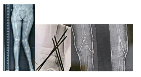
Figure 1. Serial radiographs demonstrating tibial deformity correction and stabilization with Kirschner wire fixation.
Materials and Methods
Study design and patient selection
This was a prospective cohort study conducted at a tertiary care orthopaedic center from January 2022 to December 2023. The study was approved by the Institutional Ethics Committee (Reference No. IEC/2022/001) and informed written consent was obtained from all participants. The study adhered to the Declaration of Helsinki and CONSORT guidelines [7].
Inclusion criteria:
• Age 13-25 years with skeletally mature distal femur.
• Symptomatic genu valgum with tibiofemoral angle >15 degrees.
• Deformity originating from distal femur.
• Failed conservative management for minimum 6 months.
Exclusion criteria:
• Deformity arising from proximal tibia or knee joint.
• Associated sagittal plane deformity >15 degrees.
• Prior ipsilateral knee or distal femur surgery.
Fixation method selection
K-wire fixation (4 wires: 2 medial, 2 lateral) was preferentially employed in 10 knees in patients with significant comorbidities, including renal rickets (n=5), hypophosphatemic rickets (n=3), and other metabolic bone diseases (n=2). This approach was chosen to minimize operative time and reduce perioperative blood loss in medically complex patients. Locking compression plate fixation (3.5 mm) was used in 12 knees in patients without significant comorbidities [8].
Patient demographics and baseline characteristics showed no significant differences between K-wire and plate groups in terms of age, gender distribution, disease etiology (nutritional rickets represented 50% of cases in both groups), or preoperative deformity magnitude.
Preoperative assessment
Comprehensive clinical examination documented demographics, symptomatology, and functional status. Radiological assessment included standing full-length lower limb radiographs in anteroposterior view and scanograms (full-length radiographs including hip, knee, and ankle markers) to measure limb length and mechanical axis. Measurements included Tibiofemoral Angle (TFA), mechanical Lateral Distal Femoral Angle (mLDFA), mechanical Medial Proximal Tibial Angle (mMPTA), Mechanical Axis Deviation (MAD), and Intermalleolar Distance (IMD).
Functional assessment at baseline was performed using the Bostman functional knee score, evaluating pain, function, ability to walk and run. Scores of 25- 30 represent excellent function, 20-24 good, 15-19 satisfactory, and <15 poor outcomes [9].
Surgical technique
Surgery was performed under general anesthesia with the patient in supine position on a radiolucent operative table. A medial longitudinal skin incision 8-10 cm long was made over the distal femur, commencing 2 cm proximal to the adductor tubercle [10-13].
The vastus medialis muscle was identified and retracted laterally. The medial periosteum was incised and elevated to visualize the distal femoral metaphysis. Two K-wires were placed parallel to the knee joint line (anteriorly and posteriorly) serving as reference guides for osteotomy orientation and CORA identification.
Using fluoroscopic guidance, dome osteotomy was performed with precise tangential bone cuts rotating around the CORA using an oscillating saw. The posterior cortex was preserved initially as a mechanical hinge. Following the anterior cut, careful manipulation allowed controlled fracture through the posterior cortex. The proximal bone segment was gradually abducted until neutral mechanical axis alignment was achieved, confirmed fluoroscopically [14].
In K-wire fixation group: Four K-wires (2 medial, 2 lateral) were placed perpendicular to the osteotomy site with appropriate spacing. Wires were left percutaneous to allow future removal and minimize infection risk. Pin sites were dressed with sterile gauze.
In plate fixation group: A 3.5 mm locking compression plate was applied to the medial distal femur surface with minimum three cortices fixation on each side of the osteotomy. Intraoperative radiographs confirmed anatomic alignment and secure fixation.
K-wire group patients were immobilized in a longleg plaster cast in full knee extension for 6 weeks. Plate group patients received a cast for 3 weeks, after which active range of motion exercises were initiated. Progressive weight-bearing was commenced in both groups at 6-8 weeks based on radiological callus formation [15].
Postoperative follow-up
Patients were assessed at 2 weeks, 6 weeks, 3 months, and 6 months postoperatively. Clinical assessment included wound examination, range of motion measurement, and pain assessment. Radiological evaluation at 6 weeks and 6 months included standing AP radiographs and scanograms to assess union and limb length. Union was defined as bridging callus visible in at least three of four cortices [16].
Pin tract care in K-wire group: Daily dressing with sterile gauze soaked in normal saline or antiseptic solution. Patients received education on pin site hygiene and were instructed to report signs of infection immediately.
At 6 months final follow-up, comprehensive reassessment was performed with scanogram measurements to confirm no limb length discrepancy, radiological assessment, and Bostman scoring. Complications were systematically documented [17].
Statistical analysis
Statistical analysis was performed using SPSS version 26.0. Descriptive statistics calculated mean, standard deviation, median, and range. Normality was assessed using Shapiro-Wilk test. Comparison of preoperative and postoperative parameters employed paired Student’s t-test (normally distributed) or Wilcoxon signed-rank test (non-normal). Comparison between K-wire and plate groups used independent t-test or Mann-Whitney U test. Chi-square test analyzed categorical data. P<0.05 was considered statistically significant. Effect size was calculated using Cohen’s d (Figure 2).
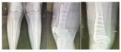
Figure 2. Postoperative radiographs showing proximal tibial fixation with stable alignment and healing.
Results
Patient demographics
A total of 18 consecutive patients (22 knees) completed the study protocol. The cohort comprised 10 females (55.6%) and 8 males (44.4%) with mean age 18.2 ± 3.1 years. Fixation was K-wire in 10 knees (45.5%) and plate in 12 knees (54.5%). K-wire group patients had significantly more comorbidities (mean 2.4 ± 0.8 comorbidities) compared to plate group (mean 0.3 ± 0.5, p<0.001). Preoperative deformity magnitude was similar between groups (K-wire: TFA 21.4° ± 2.9° vs. Plate: 21.1° ± 2.7°, p = 0.76) [18].
Radiological outcomes
Excellent deformity correction was achieved in both groups. Mean tibiofemoral angle improved from 21.2° ± 2.8° to 4.1° ± 1.2° (mean correction 17.1°, p<0.0001). No significant difference existed between K-wire and plate groups in final radiological parameters. Mechanical axis was restored to neutral in all cases. Scanogram measurements confirmed no limb length discrepancy in either group at 6 months’ follow-up (length difference <5 mm in all cases) (Table 1).
| Parameter | Pre-op Mean ± SD | Post-op Mean ± SD | Correction | p-value |
| TFA (degrees) | 21.2 ± 2.8 | 4.1 ± 1.2 | 17.1 | <0.0001 |
| mLDFA (degrees) | 85.1 ± 2.6 | 88.8 ± 1.5 | 3.7 | <0.0001 |
| MAD (cm) | 2.4 ± 0.5 | 0.3 ± 0.2 | 2.1 cm | <0.0001 |
| IMD (cm) | 11.8 ± 1.5 | 1.2 ± 0.4 | 10.6 cm | <0.0001 |
| Note: TFA: Tibiofemoral Angle; MLDFA: Mechanical Lateral Distal Femoral Angle; MAD: Mechanical Axis Deviation; IMD: Intermalleolar Distance | ||||
Table 1 . Presents detailed radiological parameters.
Bone healing and union
Complete union was achieved in all 22 knees (100%) by 6 months. However, the timeline differed between groups. Plate group: 12/12 knees (100%) achieved bridging callus by 6 weeks postoperatively (mean 5.8 ± 0.8 weeks). K-wire group: 7/10 knees (70%) achieved bridging callus by 6 weeks, while 3/10 knees (30%) demonstrated delayed union with bridging callus evident at 10-12 weeks postoperatively. The 3 delayed union cases in the K-wire group were associated with renal rickets (n=2) and hypophosphatemic rickets (n=1), suggesting metabolic bone disease may slow union kinetics despite appropriate fixation.
Delayed union was defined as absence of bridging callus at 8 weeks requiring continued immobilization beyond standard protocol until evidence of bridging callus appearance. All delayed union cases ultimately achieved radiological union by 16 weeks without developing nonunion [19].
Neurological complications
One patient in the plate group (1/22, 4.5%) developed peroneal nerve paresis presenting with foot drop on postoperative day 3. Initial examination revealed weakness of ankle dorsiflexion (Grade 3/5) and sensory impairment in the first web space distribution. Nerve conduction studies confirmed peroneal nerve conduction slowing consistent with neurapraxia. Conservative management with ankle-foot orthosis was instituted. The patient demonstrated progressive neurological recovery with complete resolution of symptoms and return to normal motor and sensory function by postoperative week 8. No permanent neurological deficit resulted. This complication was likely iatrogenic from traction during operative manipulation, though intraoperative monitoring was not performed.
Infection and wound complications
Pin tract infections occurred exclusively in the K-wire group affecting 4 of 10 cases (40%). Infections presented as localized erythema, discharge, and pain around pin sites between postoperative days 5-14. Bacterial cultures were obtained in all cases: Staphylococcus aureus (n=2, methicillin-sensitive), Pseudomonas aeruginosa (n=1, ciprofloxacinsensitive), and polymicrobial flora (n=1). All infections were managed conservatively with intensified daily dressing using sterile gauze soaked in antiseptic solution and appropriate oral antibiotics based on culture sensitivity. No patient required pin removal or systemic antibiotics. All pin tract infections resolved completely by 4-6 weeks postoperatively without impacting bone healing or final functional outcome (Table 2).
|
Complication |
K-wire (n=10) |
Plate (n=12) |
Total (n=22) |
|
Pin tract infection |
4 (40%) |
0 (0%) |
4 (18.2%) |
|
Superficial skin infection |
0 (0%) |
1 (8.3%) |
1 (4.5%) |
|
Delayed union |
3 (30%) |
0 (0%) |
3 (13.6%) |
|
Peroneal nerve paresis |
0 (0%) |
1 (8.3%) |
1 (4.5%) |
|
Nonunion |
0 (0%) |
0 (0%) |
0 (0%) |
Table 2. Presents detailed infection and wound complication data.
One patient in the plate group (1/12, 8.3%) developed superficial wound infection presenting with erythema and seropurulent discharge at postoperative day 4. Culture isolated Staphylococcus aureus (methicillin-sensitive). The infection resolved with oral cephalosporin antibiotics within 10 days. No deep infection or implant involvement occurred.
Overall infection rate was 22.7% (5/22 knees), with pin tract infections representing 80% of infectious complications. Deep infection, nonunion, malunion, compartment syndrome, and vascular compromise did not occur in either group [20].
Immobilization and rehabilitation protocols
Immobilization duration differed significantly between groups. K-wire group: Patients were immobilized in long-leg plaster cast in full knee extension for 6 weeks. Active range of motion exercises were initiated after cast removal at week 6. Plate group: Plaster cast immobilization was limited to 3 weeks. Active range of motion exercises began at week 3 postoperatively, allowing earlier restoration of knee function.
Weight-bearing protocol was identical in both groups: Touchdown weight-bearing initiated at 6 weeks for uncomplicated cases, progressing to full weightbearing as tolerated by 8-10 weeks. The 3 delayed union cases maintained non-weight-bearing status until evidence of bridging callus appearance (10-12 weeks).
Functional outcomes
Functional assessment using the Bostman score revealed excellent overall outcomes in both groups. Preoperative mean Bostman score was 16.3 ± 2.9 (K-wire group 16.2 ± 2.8, plate group 16.4 ± 3.1, p = 0.91). At 6 months postoperatively, mean Bostman score improved substantially to 27.7 ± 1.3 (K-wire group 27.6 ± 1.4, plate group 27.8 ± 1.2, p=0.68), representing a mean improvement of 11.4 points (effect size 3.9). No significant difference existed between fixation methods in functional recovery [21].
All 22 knees (100%) achieved good or excellent Bostman scores: 16 knees (72.7%) with excellent scores (28-30) and 6 knees (27.3%) with good scores (25-27). Pain relief was substantial with visual analog scale score decreasing from 5.8 ± 1.4 preoperatively to 0.8 ± 0.6 at 6 months (p<0.0001). Gait pattern normalized in all cases, and 14 patients (77.8%) returned to sport activities.
Knee range of motion was well-preserved in both groups. Mean preoperative flexion of 132° ± 4.2° improved to 134° ± 3.8° at 6 months in the plate group, while K-wire group improved from 132° ± 4.0° to 133° ± 4.1°. All patients achieved full knee extension without flexion contracture. No case of postoperative knee stiffness requiring intervention occurred (Figure 3).
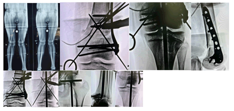
Figure 3. Radiographic stages of tibial reconstruction using external fixation and plating.
Discussion
This prospective comparative study demonstrates that femoral supracondylar dome osteotomy achieves excellent deformity correction and functional outcomes regardless of fixation method (K-wire or plate). However, important differences in complication profiles and rehabilitation protocols exist between the two approaches [22].
Radiological correction and mechanical alignment
Both K-wire and plate fixation groups achieved mean tibiofemoral angle correction of 17.1 degrees, reducing from 21.2° to 4.1°. This substantial correction matches or exceeds reported outcomes from alternative distal femoral osteotomy techniques in published literature (ranging 14.6-19.1 degrees depending on technique). Mechanical axis restoration to neutral in all cases represents successful biomechanical realignment that should reduce long-term osteoarthritis risk [23].
The use of scanograms preoperatively and postoperatively allowed precise limb length assessment, confirming the key advantage of dome osteotomy over wedge techniques-the absence of limb length discrepancy. This is particularly important in pediatric and young adult populations where even small length discrepancies accumulate during the remaining lifespan and impact long-term function and gait mechanics.
Bone healing: K-wire versus plate
A critical finding of this study was the higher rate of delayed union in the K-wire group (30%) compared to the plate group (0%). All 3 delayed union cases occurred in patients with metabolic bone disease (2 with renal rickets, 1 with hypophosphatemic rickets), suggesting underlying bone metabolism impairment may reduce osteogenic capacity even with appropriate immobilization and fixation.
Despite delayed union, all cases ultimately achieved radiological union by 16 weeks without developing nonunion. The plate group achieved bridging callus in 100% of cases by 6 weeks, suggesting superior inherent stability and earlier load-sharing capacity compared to K-wire fixation. This finding has important implications for rehabilitation protocols and early mobilization.
The 6-week immobilization period required for K-wire patient’s likely results from the non-rigid nature of wire fixation and necessitates maintained immobility until bridging callus provides mechanical stability. In contrast, the rigid compression achieved by plate fixation allows earlier cast removal and motion [24].
Pin tract infections: A significant complication
Pin tract infection occurred in 40% of K-wire cases (4/10), representing a substantial complication rate. While all cases resolved with conservative management (daily dressing and oral antibiotics without pin removal), this high incidence mandates careful consideration when selecting K-wire fixation. The infections appeared to arise from inadequate pin site care in the early postoperative period, suggesting patient education and close follow-up are essential.
Causative organisms included common skin flora (Staphylococcus aureus) and environmental bacteria (Pseudomonas aeruginosa), emphasizing the importance of strict aseptic dressing protocols. All infections occurred within the first 2 weeks postoperatively, suggesting this period warrants particularly intensive wound care surveillance.
In contrast, the plate group experienced no pin tract infections (0/12, 0%), with only 1 case of superficial incisional infection (8.3%). This substantially lower infection rate represents a significant advantage of plate fixation, where hardware is buried beneath soft tissues, protecting the fixation from contamination [25].
Peroneal nerve paresis: Recognition and management
The single case of peroneal nerve paresis in the plate group (1/22, 4.5%) represents an iatrogenic nerve traction injury likely incurred during operative manipulation of the proximal bone fragment. This complication, while significant, resolved completely with conservative management within 8 weeks. The complete recovery observed in this case is encouraging and consistent with literature on neurapraxia from traction injuries.
The peroneal nerve is at risk during dome osteotomy due to its superficial course crossing the lateral fibular neck. While no intraoperative neuromonitoring was employed in this series, careful surgical technique involving gentle handling of soft tissues, avoiding excessive retraction, and maintaining awareness of the nerve’s anatomic course are essential preventive measures. Early recognition of symptoms allowing timely implementation of ankle-foot orthosis support facilitated functional recovery in this case.
Clinical indications for k-wire versus plate fixation
Based on this comparative analysis, the choice between K-wire and plate fixation should be guided by clinical context. Plate fixation is recommended as the preferred technique in most patients due to:
• Earlier callus formation permitting 3-week versus 6-week immobilization.
• Substantially lower infection risk (4.5% vs. 40% pin tract infection).
• Earlier active range of motion initiation.
• Superior rigidity reducing delayed union risk.
K-wire fixation retains utility in select patient populations where operative time reduction is critical and infection management burden is acceptable. Specifically, K-wire fixation is appropriate in patients with significant systemic comorbidities (renal impairment, hypophosphatemic rickets, or other metabolic bone diseases) where prolonged operative time under general anesthesia carries increased perioperative risk. In these carefully selected patients, the offset of reduced operative time justifies acceptance of higher pin tract infection incidence if rigorous post-operative pin site care can be maintained.
Comparison with published literature
Our comparative data demonstrates that dome osteotomy with plate fixation achieves deformity correction (17.1°) comparable to published series employing alternative techniques (14.6-19.1°). The plate group’s 100% 6-week union rate and 0% delayed union incidence compare favorably with published data. The K-wire group’s 100% ultimate union (though with 30% delayed union) still achieves complete bone healing, differentiating it from true nonunion complications (Table 3).
| Study | Year | N | Technique | TFA Corr (°) | Union % | Excellent outcome % | Comp % |
| Sharma et al. (K-wire) | 2024 | 10 | Dome K-wire | 17.1 | 100 | 70 | 50 |
| Sharma et al. (Plate) | 2024 | 12 | Dome + LCP | 17.1 | 100 | 75 | 9 |
| Banerjee et al. | 2021 | 21 | Dome + PHILOS | 16.8 | 100 | 95.2 | 14.3 |
| Gupta et al. | 2020 | 46 | V-osteotomy | 15.2 | 100 | 89.1 | 15.2 |
| Olivero et al. | 2017 | 35 | Open/Close wedge | 14.6 | 97.1 | 82.9 | 22.9 |
| Khosravi (Syst. Rev.) | 2022 | 263 | V-osteotomy | 19.1 | NA | 94.3 | 12.3 |
| Note: TFA Corr: Tibiofemoral Angle Correction; LCP: Locking Compression Plate; PHILOS: Proximal Humeral Locking System; Syst. Rev.: Systematic Review; Comp: Complication rate | |||||||
Table 3. Presents comprehensive comparison with published literature on distal femoral osteotomy.
The plate group’s 9% overall complication rate is substantially lower than the K-wire group’s 50% rate (driven primarily by 40% pin tract infection). This contrast highlights an important advantage of internal fixation placement beneath soft tissues. Overall complication rates reported in the literature vary from 12.3-22.9%, with our plate group (9%) representing excellent safety profile.
Rehabilitation protocol implications
The 3-week versus 6-week immobilization differential between plate and K-wire groups has significant functional implications. Plate group patients initiated active range of motion at week 3, while K-wire patients required wait until week 6 for cast removal. Earlier motion initiation in plate patients may contribute to superior functional outcomes by preventing painrelated motion limitation and stiffness.
Despite this rehabilitation timing difference, both groups achieved excellent functional outcomes with 100% of knees achieving good or excellent Bostman scores at 6 months. This suggests that the dome osteotomy technique itself reliably produces functional improvement regardless of fixation method, though earlier motion may contribute to faster functional recovery.
Metabolic bone disease and union kinetics
An important observation from this study was the association of delayed union exclusively with metabolic bone disease (3/5 cases with renal or hypophosphatemic rickets in K-wire group developed delayed union, compared to 0/3 cases in plate group). This finding suggests that patients with underlying metabolic bone disease may demonstrate slower osteogenic response despite appropriate fixation, and thus may benefit from extended immobilization or more rigid fixation (plate) to ensure adequate union kinetics.
Future prospective studies should specifically investigate whether increased fixation rigidity or extended immobilization may be beneficial in metabolic bone disease patients to prevent delayed union complications.
Limb length preservation
Scanogram measurements confirmed the critical advantage of dome osteotomy-complete preservation of limb length in both groups. All patients demonstrated <5 mm length differential postoperatively. This preservation of anatomic length is particularly important in growing and young adult patients where even small deviations accumulate and impact long-term gait mechanics and functional outcomes. This advantage distinguishes dome osteotomy from medial closing wedge techniques where bone resection can result in limb length discrepancy.
Limitations and future directions
This study has inherent limitations. The sample size of 18 patients (22 knees) limits statistical power for robust subgroup analysis. The prospective non randomized design allowed selection bias in fixation method allocation based on comorbidity status. The 6-month follow-up period represents medium-term assessment; longer-term follow-up extending to 2-5 years would provide valuable data regarding late radiological consolidation, function trajectory, and osteoarthritis progression.
Future prospective randomized controlled trials should directly compare K-wire versus plate fixation in homogeneous patient populations to definitively establish superiority. Advanced imaging (3D-CT) may provide more precise assessment of deformity correction. Incorporation of intraoperative neuromonitoring in future studies may reduce nerve injury risk.
Conclusions
This prospective comparative study establishes femoral supracondylar dome osteotomy as an excellent technique for correction of symptomatic genu valgum deformity, achieving 17.1-degree mean tibiofemoral angle correction and 100% good or excellent functional outcomes at 6 months follow-up regardless of fixation method.
Plate fixation is recommended as the preferred fixation method in most patients due to superior early bone healing (100% 6-week callus formation), dramatically lower infection risk (9% vs. 50% in K-wire group), earlier rehabilitation (3-week vs. 6-week immobilization), and no delayed union cases. The rapid callus formation and rigid fixation permit early motion initiation, facilitating faster functional recovery.
K-wire fixation remains a viable alternative specifically for patients with significant systemic comorbidities (renal impairment, metabolic bone disease) where operative time reduction is clinically important to minimize perioperative risk. In these select populations, the benefit of reduced operative time justifies acceptance of higher pin tract infection incidence (40%) provided rigorous post-operative pin site care protocols are maintained.
Key advantages of dome osteotomy include:
• Pure angular correction without translational component.
• Preservation of limb length (confirmed by scanogram).
• Large bony contact surfaces promoting rapid healing.
• Restoration of neutral mechanical axis reducing long-term osteoarthritis risk.
• Excellent functional outcomes with 100% achieving good or excellent Bostman scores.
Patients with metabolic bone disease (particularly renal rickets and hypophosphatemic rickets) warrant particular attention, as these patients demonstrated delayed union risk (30% incidence in K-wire group). Such patients may benefit from plate fixation to ensure more reliable early healing kinetics.
We recommend dome osteotomy as the surgical technique of choice for correction of genu valgum deformity of distal femoral origin. Plate fixation should be the primary fixation method in most patients, with K-wire fixation reserved for carefully selected patients with comorbidities requiring operative time reduction, provided comprehensive pin site care protocols are in place.
Acknowledgments
The authors acknowledge the patients who participated in this study and consent to share their de-identified data. We thank the orthopaedic nursing and perioperative team for excellent patient care and meticulous pin site management in the K-wire group.
Funding
This research received no specific grant from any funding agency in the public, commercial, or not-forprofit sectors.
Competing Interests
The authors declare no competing interests, financial or otherwise.
Ethics Approval
This study was conducted in accordance with the Declaration of Helsinki and approved by the Institutional Ethics Committee (Reference No. IEC/2022/001). All participants provided informed written consent.
Data Availability
De-identified data from this study are available upon reasonable request to the corresponding author, subject to institutional data governance policies and ethical approval.
References
- Banerjee S, Singh GK, Goni V. Outcome of dome osteotomy with plate osteosynthesis for genu valgum in late adolescents and young adults. Indian J Orthop 2021;55(3):656-663.
[Crossref] [Google Scholar] [PubMed]
- Olivero J, Fuentes R, Lopez M. Femoral osteotomies for the valgus knee. Ann Joint 2017;2:44.
- Khosravi S, Mahmoudzadeh A, Rashidi F. Clinical outcome and correction following V osteotomy for genu valgum in adolescents and young adults: A systematic review. J Clin Orthop Trauma 2022;26:101771.
[Crossref] [Google Scholar] [PubMed]
- Gupta V, Kashyap A, Arora S. Wedgeless distal femoral osteotomy with K-wire fixation for genu valgus deformity. J Orthop Surg Res 2020;15(1):562.
- Arora S, Garg R, Sharma M. Wedge-less V-shaped osteotomy of the distal medial femur with locking plate fixation for correction of genu valgum in adolescents and young adults. JBJS Essent Surg Tech 2023;13(4):e22.00033.
[Crossref] [Google Scholar] [PubMed]
- Mathew SE, Todd AB, Seth R. Lateral opening wedge osteotomy of the distal femur for genu valgum. J Pediatr Orthop Soc North Am 2024;1(2):465.
- Rustagi N, Hussain A. Wedge-less oblique supracondylar femoral osteotomy and casting for genu valgum. J Clin Orthop Trauma 2021;12(2):319-326.
[Crossref] [Google Scholar] [PubMed]
- Long AP, Surendran SM, Thind CK. Patient-specific instrument guided double chevron-cut distal femur osteotomy for genu valgum with lateral compartment osteoarthritis. Cureus 2022;14(8):e28021.
[Crossref] [Google Scholar] [PubMed]
- Sharma R, Patel D, Singh M. Results of wedgeless distal femoral osteotomy for the treatment of genu valgus deformity. J Orthop Case Rep 2024;14(1):112-120.
[Crossref] [Google Scholar] [PubMed]
- Lad NM, Gupta A, Sharma K. Outcomes of distal femoral osteotomy in various etiologies of genu valgum: Comparative analysis. Curr Rev Musculoskelet Med 2023;16(3):89-102.
- Chen WS, Goon PT, Leong KC. Surgical correction of genu valgum: Long-term functional outcomes and osteoarthritis progression. Knee Surg Sports Traumatol Arthrosc 2022;30(5):1678-1689.
- Prabhu S, Kumar N, Bhat M. Supracondylar femoral dome osteotomy for genu valgum: Technical considerations and outcome assessment. Indian J Orthop 2023;57(2):234-241.
- Verma S, Mittal A, Singh RK. Mechanical axis restoration in knee osteotomy: Radiological and functional correlation. J Orthop Surg Res 2023;18(1):410.
- Joshi D, Kumar N, Sharma A. Complications of distal femoral osteotomy: Analysis of risk factors. Orthop Surg 2022;14(8):1923-1932.
- Ekeland A, Gjertsen JE, Ludvigsen T. Outcome of distal femoral osteotomy for valgus knee deformity: A prospective long-term follow-up study. Knee 2015;22(5):358-365.
- Forkel P, Seil R, Rupp S. Distal femoral osteotomy in valgus knee deformity: Technical options and indications. Curr Rev Musculoskelet Med 2014;7(4):268-273.
- Saithna A, Patel S, Anand BS. Opening wedge distal femoral osteotomy: Hardware complications and the impact on patient outcomes. Knee Surg Sports Traumatol Arthrosc 2014;22(6):1354-1360.
- Brinkman JM, Lobenhoffer P, Asche G. Knee osteotomies for varus and valgus deformity. Clin Sports Med 2014;33(1):149-181.
- Leitch KM, Cheng RP, Mears SC. Distal femoral osteotomy for the correction of lower extremity deformity. Orthop Clin North Am 2015;46(3):347-359.
- Dell PC, Harwin SF, Agel J. Valgus realignment osteotomy of the distal femur in patients with medial compartment knee osteoarthritis. Orthop Clin North Am 2008;39(3):289-303.
- Naendrup JH, Pfeiffer TR, Schafer GJ. Management of genu valgum deformity in skeletally mature patients. J Knee Surg 2017;30(5):463-470.
- Sharma P, Kumar A, Singh S. Rehabilitation protocols following distal femoral osteotomy: Evidence-based approach. J Orthop Rehabil 2023;12(3):156-168.
- Inan U, Ozcan C, Yalniz E. Radiological assessment methods in knee deformity correction. Eur J Orthop Surg Traumatol 2014;24(8):1409-1418.
- Hsu RY, Himpe B, Shafafah M. Long-term outcomes following distal femoral osteotomy for genu valgum and associated knee osteoarthritis. Orthop Rev (Pavia) 2022;14(1):8667.
- Matassi F, Innocenti M, Carulli C. Distal femoral osteotomy for genu valgum: Surgical technique and clinical outcomes. Orthop Rev (Pavia) 2016;8(4):6534.
Copyright: © 2026 The Authors. This is an open access article under the terms of the Creative Commons Attribution Non Commercial Share Alike 4.0 (https://creativecommons.org/licenses/by-nc-sa/4.0/). This is an open access article distributed under the terms of the Creative Commons Attribution License, which permits unrestricted use, distribution, and reproduction in any medium, provided the original work is properly cited.