Research Article - Archives of Clinical and Experimental Surgery (2025)
Increased Stretched Penile Length in Vesical Calculus: A Study of 50 Paediatric Cases
Ashok Kumar Ladda, Maneesh Kumar Joleya*, Pooja Tiwari, Vinod Raj, Shashi Shankar Sharma, Ram Mohan Shukla, B.K. Lahoti and Manoj JoshiManeesh Kumar Joleya, Department of Department of Pediatric Surgery, SSH and MGMMC, Indore, India, Email: drjollymaneesh@yahoo.co.in
Received: 27-Jan-2025, Manuscript No. EJMACES-25-160071; Editor assigned: 30-Jan-2025, Pre QC No. EJMACES-25-160071 (PQ); Reviewed: 13-Feb-2025, QC No. EJMACES-25-160071; Revised: 15-Apr-2025, Manuscript No. EJMACES-25-160071 (R); Published: 13-May-2025
Abstract
Background: Primary vesical calculus is common among Indian males aged 1 to 10 years. Parents/guardians can report that the child cries during micturition and often pulls at his penis to relieve pain. Repeated pulling/tugging/massaging and ensuing penile erection increases penile smooth muscles’ exposure to stretch. Prolonged semi erection in high flow priapism has demonstrated increased penile lengths in prepubescent boys. We assess whether pediatric vesical calculus cases demonstrate a longer penile length compared to controls and determine the contributions of age, duration of pulling, and stone width towards penile length.
Methods: Cross-sectional study tertiary care center. We consecutively included 50 ultrasonographically confirmed pediatric vesical calculus cases and 150 controls. Stretched penile length was measured preoperatively and stone width postoperatively following percutaneous cystolithotomy. Histories on duration of pulling of penis were queried. Statistical analysis will ascertain if significant difference exists between stretched penile lengths of cases and controls. Contributions from duration of pulling and stone width will be examined using correlations and univariate followed by multivariate regressions.
Results: Independent t-test reveal higher stretched penile lengths among cases compared to controls (T=6.412, p<0.001). Stretched penile length positively correlated with age (ρ=0.274, p=0.053), duration of pulling (ρ=0.511, p<0.001), and stone width (r=0.364, p=0.009). Multivariate polynomial regression utilizing age, duration of pulling, and stone width yields an adjusted R-squared of 0.3181 (p=0.006).
Conclusion: Increased stretched penile length constitutes a diagnostic pearl for vesical calculus in children which can ensure timely referral and intervention for preventing complications in rural settings lacking imaging modalities and specialists.
Keywords
Bladder stone; Pediatric; Penile length; Stretched penile length; Vesical calculus; Level of evidence
Abbreviations
VC: Vesical Calculus; SPL: Stretched Penile Length; PTM: Pulling/Tugging/ Massaging; dPTMp: duration of Pulling/Tugging/Massaging the penis; MWoS: Maximum Width of Stone; SAC: Stretch Activated Channels; VDCC: Voltage Dependent Calcium Channel; fPTMp: Frequency of Pulling/Tugging/Massaging the penis
Introduction
Primary vesical Calculus (VC) is a common disease in children. The incidence of urinary bladder calculi has decreased to a great extent, owing to nutritional improvement in the diet among different populations. A broad geographic belt encompassing Southeast Asia, northern Africa, the Middle East, and the Indian subcontinent are regions where incidence of VC is higher in children, predominant in ages 1.5 to 14 years and males [1]. In developing nations, VC are common in children owing to risk factors like low dietary intake of phosphate, vitamin A deficiency, dehydration, infection, and low protein diet. Adults with VC can present asymptomatically or with symptomatology including dysuria, suprapubic pain, weak urinary stream, sudden voiding stoppage, terminal haematuria, and varying degrees of pain present on the tip of the penis, scrotum, pelvis, or the perineum [2]. Among pediatric population, the child or the parents/guardians will complain difficulty in micturition, urinary retention, increased frequency, recurrent episodes of diarrhoea, and recurrent urinary tract infections with rarer histories of fever, family history of stone disease, and even rectal prolapse [1]. Parents/guardians also complain that the child cries every time he passes urine and pulls/ tugs/massages his penis in an attempt to relieve the pain, and oftentimes passes blood in urine [3,4]. Bladder may be distended, and the foreskin may be red and swollen from being pulled on; the examiner may be able to feel the stone on a rectal examination. Although the stone is likely a urate stone, it will probably contain enough calcium for one to see it on an X ray. Isolated to a previous case report, children with VC had demonstrated an abnormal increase in penile length [4]. This was attributed to repeated pulling and stretching of the penis creating a prolonged state of semi erection which has previously demonstrated to contribute to an increase in the penile length in prepubescent boys with high-flow priapism [5].
This is an interesting clinical observation warranting the conduct of this study. The primary objectives of the present study is to:
• To ascertain if there is significant difference between Stretched Penile Lengths (SPL) in pediatric patients of VC compared to normal pediatric individuals in the corresponding age group (2-10 years).
• To ascertain the correlation between SPL and Pulling/Tugging/Massaging (PTM) episodes i.e., duration of PTM the penis (dPTMp), and between SPL and the Maximum Width of the Stone (MWoS).
• Perform a multivariable regression to ascertain the determinants which impact the SPL in a pediatric patient of VC.
dPTMp measures the duration since exposure to mechanical stretch due to PTM and mechanical stretch ensuing from erection or semi erection due to PTM of the penis. Secondary objectives include assessing the correlation between each of SPL, dPTMp, and MWoS with age of children with VC, between SPL of the control population and age, and between dPTMp and MWoS.
Materials and Methods
Study design
Cross sectional study. We will be including a total of 50 consecutive pediatric participants with ultrasonography confirmed VC as cases/subjects in our study. Three pediatric controls will be matched for each case according to age i.e., total 150 individuals will be considered to form the control population. Guardians will be explained about the nature of study design the child is considered for and informed consent will be obtained for individual cases and controls prior to enrolment. Ethical approval was obtained to conduct the study from the local institutional ethics committee. The study was conducted in accordance with the code of ethics of the World Medical Association (Declaration of Helsinki). Demographic details and history on dPTMp will be queried and captured. If dPTMp cannot be recalled with certainty, it will be recorded as “Not available” and excluded from analysis. SPL measurements will be taken according to the Methods section described below for both cases and controls. Morphological characteristics of VC or MWoS will be recorded postoperatively following suprapubic cystolithotomy. Duration of study lasted from February 2013 to February 2016.
Inclusion criteria for cases
• Males diagnosed with vesical calculus.
• Age between 2 to 10 years.
• Admitted in the pediatric surgery division of Maharaja Yashwantrao (MY) Hospital, Indore from February 2013 to February 2016.
Rationale for the inclusion criteria: Tomova, et al., conducted a cross-sectional study of 6200 males [6]. The following were the findings:
• The testes begin to increase in size at the ages 8 to 9 years. Before the onset of puberty, the growth of testes is minimal.
• The increase in penile length is significant only after one year of increase in size of testes.
• Although penile length and circumference demonstrated gradual growth, the period of maximum growth was approximately 12 to 16 years of age.
Until age 10 years, the mean penile length remained less than 5 cm. This forms the rationale why we have chosen the age group 2-10 years as subjects for our study.
Inclusion criteria for controls
• Admitted within the same MY Hospital pediatric surgery unit for conditions other than urogenital problems.
• Participants belong to geographical area covered by the same tertiary health centre (i.e., MY hospital, Indore).
Exclusion criteria for cases and controls
• Associated congenital genital anomalies (like hypospadias, epispadias).
• Presence of secondary sexual characters.
• Associated primary skin diseases involving the perineal region or the penile skin which can impact pain symptomatology and its effect on SPL or interferes with measurement of SPL.
Methods
SPL is defined as the linear distance along the dorsal side of the penis extending from the pubo-penile skin junction to the tip of the glans with penis stretched longitudinally in flaccid state. Measurements will be taken three times and by a single observer to mitigate any technical error during measurement. Measurement will be considered till tip of glans; prepuce skin is not taken into consideration. Measurement will be recorded prior to foley’s catheterization. Measurement will be recorded up to one point after decimal.
Statistical analysis
For comparison of continuous data between cases and controls, independent sample t-test will be utilized for normally distributed data and Mann Whitney U test/Wilcoxon signed rank test for non-normal distribution. Correlations will be assessed using Pearson’s correlation coefficient and Spearman’s correlation coefficient for normally and non-normally distributed data respectively. Univariate regression will be run for age, dPTMp, and MWoS individually to assess their impact on SPL and statistically significant parameters will be considered for multivariable regression. A p-value ≤ 0.05 was considered as the statistical significance threshold for all the above analyses.
Results
We examined a total of 50 cases from ages 2 to 10 years (Mean=5.32 years ± 2.40) (Table 1). dPTMp was available for 37 cases ranging from 1 day to 36 months (Mean=10.44 months ± 10.478), while MWoS was available for all 50 cases ranging from 0.5 cm to 6 cm (Mean=2.29 cm ± 1.112). The mean SPL of cases was found to be 6.09 cm ± 1.534 (n=50) and of controls was 4.69 cm ± 0.292 (n=150) (Table 2). As the SPL was near normally distributed, independent sample t-test was utilized. A t-statistic of 6.412 was obtained which demonstrated a significant difference between SPLs of cases and controls which was also statistically significant (p<0.001) with SPLs being higher among cases.
| Sl. no. | Age (in years) | Stretched penile length (in cm) cases | Stretched penile length (in cm) controls | History of pulling/tugging/massaging of the penis |
Duration of pulling/tugging/massaging of the penis (in months) | Maximum width of stone (in cm) |
|---|---|---|---|---|---|---|
| 1 | 2 | 11 | 4.2, 4.3, 4.6 | Yes | 12 | 2.5 |
| 2 | 2 | 6 | 4.3, 4.1, 4.6 | No | NA | 1 |
| 3 | 2 | 5.4 | 4.5, 4.6, 4.3 | Yes | 2 | 1.5 |
| 4 | 2 | 6.2 | 4.4, 4.3, 4.5 | Yes | 2 | 1.5 |
| 5 | 2 | 4.6 | 4.3, 4.4, 4.4 | No | NA | 1.5 |
| 6 | 2 | 4.5 | 4.3,4.6, 4.5 | No | NA | 3.3 |
| 7 | 3 | 7 | 4.6, 4.2, 4.0 | Yes | 6 | 2 |
| 8 | 3 | 7 | 4.7, 4.9, 4.7 | Yes | 3 | 2 |
| 9 | 3 | 5 | 4.5, 4.3, 4.4 | Yes | 0.6 | 1 |
| 10 | 3 | 6 | 4.1, 4.2, 4.3 | Yes | 3 | 2 |
| 11 | 3 | 7 | 4.6, 4.7, 4.5 | Yes | 6 | 2 |
| 12 | 3 | 4.5 | 4.0, 4.8, 4.5 | Yes | 0.5 | 1 |
| 13 | 3 | 5.8 | 4.4, 4.5, 4.6 | Yes | 6 | 2.2 |
| 14 | 3 | 5.8 | 4.5, 4.4, 4.7 | Yes | 3 | 2 |
| 15 | 4 | 5 | 4.4, 4.1, 4.4 | Yes | 0.1 | 1 |
| 16 | 4 | 5 | 4.1, 4.5, 4.6 | Yes | 8 | 2.5 |
| 17 | 4 | 5.5 | 4.1. 4.3, 4.5 | No | NA | 1.5 |
| 18 | 4 | 6 | 4.4, 4.7, 4.9 | No | NA | 3 |
| 19 | 4 | 3 | 4.6, 4.6, 4.5 | Yes | 3 | 1.5 |
| 20 | 4 | 5.3 | 4.7, 4.5, 4.7 | No | NA | 2 |
| 21 | 5 | 6.5 | 4.5, 4.6, 4.6 | Yes | 6 | 2 |
| 22 | 5 | 4.8 | 4.8, 4.3, 4.6 | Yes | 1 | 1.2 |
| 23 | 5 | 6.8 | 4.5, 4.4, 4.5 | Yes | 6 | 3 |
| 24 | 5 | 4.5 | 4.5, 4.5, 4.6 | No | NA | 0.5 |
| 25 | 5 | 6.8 | 4.7, 4.5, 4.6 | Yes | 10 | 2 |
| 26 | 5 | 6.8 | 4.6, 4.7, 4.4 | Yes | 1 | 2.5 |
| 27 | 5 | 7.6 | 4.4, 4.4, 4.5 | Yes | 12 | 3.5 |
| 28 | 5 | 6.2 | 4.8, 4.6, 4.5 | No | NA | 2 |
| 29 | 5 | 6 | 4.8, 4.5, 4.7 | Yes | 4 | 2 |
| 30 | 5 | 4.6 | 4.5, 4.6, 4.8 | No | NA | 1.2 |
| 31 | 6 | 6.3 | 4.5, 4.7, 5.0 | Yes | 12 | 4 |
| 32 | 6 | 4.9 | 5.0, 4.8, 4.7 | No | NA | 1 |
| 33 | 6 | 4 | 4.8, 4.9, 5.1 | No | NA | 2 |
| 34 | 6 | 6.4 | 5.0, 4.7, 5.0 | Yes | 16 | 2.2 |
| 35 | 7 | 12 | 4.9, 5.0, 5.0 | Yes | 24 | 2.6 |
| 36 | 7 | 6 | 4.5, 5.2, 5.1 | Yes | 24 | 3 |
| 37 | 7 | 5 | 4.6, 5.0, 5.0 | No | NA | 2 |
| 38 | 7 | 4.7 | 4.9, 5.0, 4.8 | Yes | 24 | 1.7 |
| 39 | 7 | 5.9 | 5.1, 5.0, 4.7 | Yes | 36 | 2 |
| 40 | 7 | 5.9 | 4.9, 4.9, 5.1 | Yes | 7 | 2 |
| 41 | 8 | 5.4 | 4.9, 4.9, 4.9 | No | NA | 6 |
| 42 | 8 | 5.9 | 4.9, 4.8, 4.9 | Yes | 1 | 2 |
| 43 | 8 | 6 | 5.0, 4.9, 5.0 | Yes | 1 | 3 |
| 44 | 8 | 6 | 4.7, 4.9, 4.6 | Yes | 9 | 2.4 |
| 45 | 9 | 6.5 | 5.0, 5.1, 5.0 | Yes | 12 | 3 |
| 46 | 9 | 7.2 | 5.3, 5.0, 5.2 | Yes | 14 | 3.2 |
| 47 | 10 | 7.6 | 5.0, 5.3, 5.1 | Yes | 36 | 3 |
| 48 | 10 | 7.5 | 5.4, 5.3, 5.4 | Yes | 36 | 6 |
| 49 | 10 | 7 | 5.0, 5.1, 5.0 | Yes | 24 | 2 |
|
50 |
10 |
8.3 |
5.0, 5.1, 5.1 |
Yes |
15 |
4.5 |
Table 1. Data collected: Age, stretched penile length, mean stretched penile length, history of pulling if the penis, duration of pulling and maximum width of stone.
| Group statistics | ||||||
|---|---|---|---|---|---|---|
| Group | N | Mean | Std. deviation | T | p-value | |
| Stretched penile length (cm) | Case | 50 | 6.086 | 1.5340 | 6.412 | <0.001 |
| Control | 150 | 4.687 | 0.2921 | |||
Table 2. Independent t-test between case and control.
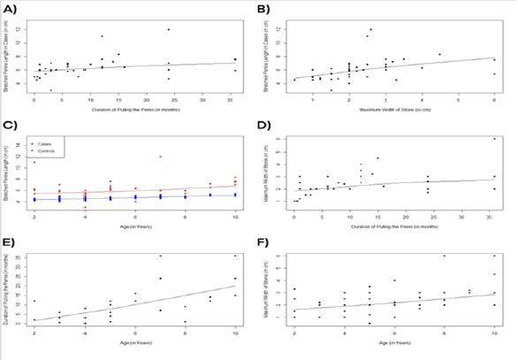
SPL demonstrated moderate positive correlation with both dPTMp (ρ=0.511, p<0.001) and MWoS (r=0.364, p=0.009) both of which were statistically significant. SPL additionally demonstrated a weak positive correlation with age of children with VC (ρ=0.274, p<0.001) and a strong positive correlation with age of control population (ρ=0.779, p<0.001). dPTMp (ρ=0.597, p<0.001) and MWoS (ρ=0.459, p<0.001) positively correlated with age of children with VC. MWoS moderately and positively correlated with dPTMp (ρ=0.605, p<0.001) indicating that it can serve as a surrogate to dPTMp in determining SPL (Table 3 and Figure 1).
| Independent variable and dependent variable | Correlation test used | Correlation coefficient | p-value |
|---|---|---|---|
| Stretched Penile Length in cases (in cm) and duration of pulling of penis (in months) (n=37) | Spearman's rank correlation | 0.511 | p<0.001 |
| Stretched penile length in cases (in cm) and maximum width of stone (in cm) (n=50) | Pearson correlation coefficient | 0.364 | p=0.009 |
| Stretched penile length in cases (in cm) and age (in years) (n=50) | Spearman's rank correlation | 0.274 | p=0.053 |
| Stretched penile length in controls (in cm) and age (in years) (n=150) | Spearman's rank correlation | 0.779 | p<0.001 |
| Maximum width of stone (in cm) and duration of pulling of penis (in months) (n=37) | Spearman's rank correlation | 0.605 | p<0.001 |
| Duration of pulling of penis (in months) and age (in years) (n=37) | Spearman's rank correlation | 0.597 | p<0.001 |
| Maximum width of stone (in cm) and age (in years) (n=50) | Spearman's rank correlation | 0.459 | p<0.001 |
Table 3. Correlations.
Figure 1. Scatter plots and loess lines demonstrating the relationship between A) SPL in cases (in cm) and dPTMp (in months); B) SPL in cases (in cm) and MWoS (in cm); C) SPL in cases (red) and controls (blue) (in cm) and Age (in years); D) MWoS (in cm) and dPTMp (in months); E) dPTMp (in months) and Age (in years); F) MWoS (in cm) and Age (in years).
Scatter plots with loess lines demonstrated a polynomial variation of SPL with age, dPTMp, and MWoS. Univariate polynomial regression demonstrated that age (Adjusted R-squared=0.07539, p=0.059), dPTMp (Adjusted R-squared=0.2056, p=0.007), and MWoS (Adjusted R-squared=0.212, p=0.00139) yielded a higher adjusted R-squared compared to their corresponding univariate linear regressions with only dPTMp and MWoS reaching statistically significance. In light that age did ensue in a higher adjusted R-squared and higher statistical significance with polynomial regression trials compared to linear, we decided to consider age in the multivariate regression. Multivariate polynomial regression utilizing age, dPTMp, and MWoS revealed a higher adjusted R-squared of 0.3181 which was highly statistically significant (p=0.006) compared to multivariate linear regression.
Discussion
Urinary bladder calculi can be primary or secondary. Primary bladder calculi form in sterile urine without any known underlying anatomical defect and are common in children younger than the age of 10, with a peak incidence at 2 to 4 years of age. The disease is much more common in boys than in girls, with ratios ranging from 9:1 to as high as 33:1 in certain areas of India. Secondary bladder calculi are common in adults and are manifestations of an underlying pathologic condition like urethral strictures, benign prostatic hyperplasia, bladder neck contractures, flaccid or spastic neurogenic bladders and foreign bodies [2,3,7].
The bladder derives its somatic innervation from the nucleus of Onuf/sacral pudendal nucleus in the ventral horn via the pudendal nerve. The nerves to the penis are derived from the pudendal and cavernous nerves. The pudendal nerves supply somatic motor and sensory innervation to the penis. Therefore, in patients with vesical calculus, pain is often referred to the tip of the penis. Young boys with a bladder stone are known to repeatedly pull and squeeze the penis in a futile attempt of alleviating pain [3]. Physical stretching of the penis or tumescence caused by repeated massage could be responsible for lengthening of the penis. This hypothesis is supported by a recent publication wherein prolonged semi erection of high flow priapism in a prepubertal boy was shown to increase penile size [5].
In this study, we demonstrated a higher SPL among pediatric patients with vesical calculus compared to controls which is statistically significant. We demonstrated that SPL in pediatric patients of vesical calculus correlates positively with age, dPTMp, and MWoS. To understand basis for this finding, it is imperative to understand the impact of stretch stimulus on smooth muscles. The penis is formed by three cylindrical masses of erectile tissue, two corpora cavernosa and one corpora spongiosum, the former being larger. They contain interconnected caverns or vascular spaces which when ultimately filled with blood will confer a tumescent state to the penis. The vascular spaces are enclosed by interwoven trabeculae composed of mainly smooth muscles and connective tissue. Repeated PTM of the penis alone or together with the tumescent state conferred introduces repeated and persistent stretching of these smooth muscles. Persistent stretch stimulus on smooth muscles due to distension or pressure leading to hypertrophic changes within myocytes has been demonstrated in multiple organs including bladder, intestine, blood vessels, ureter, and bile duct, in both animal models and humans [8-13]. Portal vein organ cultures demonstrate a longitudinal stretch dependent enhanced synthesis of smooth muscle α-actin, actin-binding proteins (SM22α and calponin), and the intermediate filament protein desmin, all of which are suggestive of smooth muscle hypertrophy [9]. In portal vein smooth muscles, stretch induces focal adhesion kinase phosphorylation with an acute phase (<15 minutes) comprising of MAPK pathway activation and a long term (>24 hours) phase correlating with Rho/Rho Kinase pathway activation. In a study by Richard. et al., [8] constant mechanical stretch for a duration of 48 hours demonstrated smooth muscles actively entering cell division resulting in a time-dependent smooth muscle hypertrophy along with hyperplasia which was MAPK-dependent. Downregulation of MAPK pathway inhibits stretch induced smooth muscle hypertrophy, hyperplasia, and nuclear protein import. Additional mechanisms of stretch-induced smooth muscle growth include both voltage-dependent and independent increases in intracellular calcium levels (Ca2+) [9,10,12,14]. Stretch Activated Channels (SAC) induce sustained membrane depolarization via cation influx (mainly Na+) and activates calcium entry through Voltage Dependent Calcium Channel (VDCC) into the smooth muscle cells and enhance cytosolic calcium [9]. Calcium is required for the synthesis of smooth muscle-specific proteins which is driven by VDCC-mediated Rho/ROK activation which through myocyte enhancer factor 2A and 2D regulates myocardin expression and actin polymerization. Voltage-independent mechanisms also include the activation of stretch activated channels which have been found in airways, bladder, gastrointestinal tract, and vascular smooth muscles and can have different ionic permeabilities (Ca2+, Cl-, K+ or Na+) [9,10,15,16]. In addition to increase in cytosolic calcium through entry via Ca2+ specific SAC, cytosolic calcium levels are amplified through calcium release from sarcoplasmic reticulum. These findings can explain the foundational mechanisms of proliferation and growth in erectile tissue smooth muscles of the penis exposed to repeated and persistent stretching from PTM or persistent tumescence ultimately leading to increase in the penile length.
Polynomial regressions run for each of age, dPTMp and MWoS demonstrated a higher adjusted R-squared and higher statistical significance compared to corresponding univariate linear regressions. All three determinants were considered for multivariate polynomial regression and correspondingly demonstrated a higher adjusted R-squared and higher statistical significance compared to multivariate liner regression. While it is the most robust model utilizing current determinants, they explain for only 31.81% of the model and a possible limitation lies with using dPTMp as a determinant. dPTMp is subject to recall bias and social desirability bias which can impact the histories and longer duration may not necessarily imply longer exposure to stretch stimuli. Frequency of PTM the penis (fPTMp) would be a better and robust determinant of total stretch exposure than dPTMp. Categorizing frequency and/or duration histories on Likert Scales can mitigate the impact of recall and social desirability bias allowing for an accurate estimation stretch exposure. We additionally demonstrated that MWoS positively correlated with dPTMp which can be explicable considering the longer a stone is retained in the bladder, additional concentric layers of stone material will be deposited until they are too large to pass and become symptomatic and hence, MWoS can serve as an alternative determinant of SPL for further studies [2].
A major limitation of this study is that it assumes a causative nature of PTM episodes towards increased SPL measurements. Lack of predictors of vesical calculi prevent their early detection, and particularly in this study, prior to any PTM episodes. For this reason, we could not capture pre-PTM episode SPL measurements objectively or even subjectively when parents/guardians were queried to describe in laymen terms of any relative size change before and after the PTM episodes. This allows us to derive two inferences. First, if a causative nature is true, our study design and the validity of results holds. Or, secondly, if SPL measurements are independent of PTM episodes, this implies that the cases in our study had physiologically larger SPLs even prior to developing vesical calculi and PTM of the penis. Can this propose that physiologically larger SPLs are associated with a higher risk of developing vesical calculi i.e., is it a risk factor? While the study could not capture this, it will be a difficult task even for further attempts exploring this finding considering the lack of pragmatic opportunities to record pre-PTM SPLs in the absence of screening recommendations.
Another limitation of this study includes an inability to directly capture the intensity and duration of penile tumescence and the impact of tumescent stretch on SPL. Eliciting histories on percentage of PTM episodes associated with tumescence and their duration form crucial determinants of SPL in VC patients than PTM stretch alone. We recommend a combination of fPTMp, MWoS, and tumescence variables in further studies estimating SPL in pediatric patients of VC. However, maintaining a sensitive context while performing these enquiries, parent/guardian uncomfortableness or awkwardness with the topic, social desirability bias and a child’s or guardians’ ability to understand and provide an accurate history can form the limitations with using tumescence as a determining factor for SPL.
Conclusion
In conclusion, this study reports higher SPL in pediatric patients of VC compared to normal pediatric population. This finding aids purely as a diagnostic sign for VC in children from resource limited settings where availability of imaging modality and specialists is a concern. Timely referral to tertiary centres and intervention can prevent complications of infection. We also provided additional parameters which can be surveyed in further studies of vesical calculus in pediatric and/or adult populations which can prove to be more robust.
References
- Lal B, Paryani JP. Childhood bladder stones-an endemic disease of developing countries. J Ayub Med Coll Abbottabad 2015;27(1):17-21.
[Google Scholar] [PubMed]
- Leslie SW, Sajjad H, Murphy PB. Bladder Stones. In: StatPearls [Internet]. Treasure Island (FL): StatPearls Publishing. 2022,994.
- Marley J. Campbellâ?Walsh Urology, (Edition)â?Edited by AJ Wein, LR Kavoussi, AC Novick, AW Partin and CA Peters.
- Raveenthiran V. Abnormally long penis in children with vesical calculus. Indian J Surg 2013;75(1):62-63.
[Crossref] [Google Scholar] [PubMed]
- Awwad ZM. Does prolonged semi erection in prepubertal high flow priapism result in increased penile size? Saudi Medical J 2005;26(3):481-483.
[Google Scholar] [PubMed]
- Tomova A, Deepinder F, Robeva R, Lalabonova H, Kumanov P, Agarwal A. Growth and development of male external genitalia: A cross sectional study of 6200 males aged 0 to 19 years. Arch Pediatr Adolesc Med 2010;164(12):1152-1157.
[Crossref] [Google Scholar] [PubMed]
- Stoller ML, Bolton DM. Chapter 17 Urinary stone disease. Smith and TanaghoÂs general urology 18e, Lange Medical Books/McGraw-Hill Companies, Inc. 2013.
- Richard MN, Deniset JF, Kneesh AL, Blackwood D, Pierce GN. Mechanical stretching stimulates smooth muscle cell growth, nuclear protein import and nuclear pore expression through mitogen activated protein kinase activation. J Biol Chem 2007;282(32):23081-23088.
[Crossref] [Google Scholar] [PubMed]
- Ren J, Albinsson S, Hellstrand P. Distinct effects of voltage-and store-dependent calcium influx on stretch-induced differentiation and growth in vascular smooth muscle. J Biol Chem 2010;285(41):31829-31839.
[Crossref] [Google Scholar] [PubMed]
- Sweeney M, Yu Y, Platoshyn O, Zhang S, McDaniel SS, Yuan JX. Inhibition of endogenous TRP1 decreases capacitative Ca2+ entry and attenuates pulmonary artery smooth muscle cell proliferation. American J Physiol Lung Cell Mol Physiol 2002;283(1):144-155.
[Crossref] [Google Scholar] [PubMed]
- Gabella G. Hypertrophy of visceral smooth muscle. Anat Embryol 1990;182(5):409-424.
[Crossref] [Google Scholar] [PubMed]
- Sigurdson W, Ruknudin A, Sachs F. Calcium imaging of mechanically induced fluxes in tissue-cultured chick heart: Role of stretch-activated ion channels. Am J Physiol Heart Circul Physiol 1992;262(4):1110-1115.
[Crossref] [Google Scholar] [PubMed]
- Yamaguchi O. Response of bladder smooth muscle cells to obstruction: Signal transduction and the role of mechanosensors. Urology 2004;63(3):11-16.
[Crossref] [Google Scholar] [PubMed]
- Kirber MT, Guerreroâ?Hernández A, Bowman DS, Fogarty KE, Tuft RA, Singer JJ, et alS. Multiple pathways responsible for the stretch induced increase in Ca2+ concentration in toad stomach smooth muscle cells. J physiol 2000;524(1):3-17.
[Crossref] [Google Scholar] [PubMed]
- Guibert C, Ducret T, Savineau JP. Voltage-independent calcium influx in smooth muscle. Progress Biophy Mol Biol 2008;98(1):10-23.
[Crossref] [Google Scholar] [PubMed]
- Bergdahl A, Gomez MF, Wihlborg AK, Erlinge D, Eyjolfson A, Xu SZ, et al. Plasticity of TRPC expression in arterial smooth muscle: Correlation with store-operated Ca2+ entry. Am J Physiol Cell Physiol 2005;288(4):C872-C880.
[Crossref] [Google Scholar] [PubMed]
Copyright: © 2025 The Authors. This is an open access article under the terms of the Creative Commons Attribution Non Commercial Share Alike 4.0 (https:// creativecommons.org/licenses/by-nc-sa/4.0/). This is an open access article distributed under the terms of the Creative Commons Attribution License, which permits unrestricted use, distribution, and reproduction in any medium, provided the original work is properly cited.


