
 +44-74-1148-3554




Research Article - Archives of Clinical and Experimental Surgery (2026)
Lateral Column Shortening with Talonavicular Fusion and Adductor Hallucis Lengthening in the Management of Neglected and Relapsed Congenital Talipes Equinovarus: A Retrospective Analysis of 22 Cases
Manish Bajaj*, Gaurav Jain and Anshul MeenaManish Bajaj, Department of Orthopaedic Surgery, LNCT Medical College, Indore, India, Email: manishbajaj@gmail.com
Received: 15-Jun-2026, Manuscript No. EJMACES-26-192787; Editor assigned: 17-Jun-2026, Pre QC No. EJMACES-26-192787 (PQ); Reviewed: 01-Jul-2026, QC No. EJMACES-26-192787; Revised: 10-Jul-2026, Manuscript No. EJMACES-26-192787 (R); Published: 17-Jul-2026
Abstract
Background: Neglected and relapsed Congenital Talipes Equinovarus (CTEV) presents significant challenges in pediatric orthopedic surgery. While the Ponseti method has revolutionized initial management, many patients still require surgical intervention for persistent or recurrent deformities. This study evaluates the efficacy of combined lateral column shortening, talonavicular fusion, and adductor hallucis lengthening in managing resistant deformities.
Methods: A retrospective analysis was conducted on 22 patients (34 feet) with neglected or relapsed CTEV who underwent lateral column shortening with cuboid wedge resection, talonavicular fusion, and adductor hallucis lengthening between 2015 and 2022. Patients had a minimum follow-up of 24 months. Clinical assessment included morphological evaluation, functional outcomes, and radiographic analysis using standardized measurements. Paired t-tests and chi-square analysis were employed (p<0.05 considered significant).
Results: Mean patient age at surgery was 8.4 ± 3.2 years. Mean follow-up duration was 34.6 ± 8.4 months. Preoperative talus-first metatarsal angle (AP view) averaged 35.2° ± 8.6°, improving to 18.3° ± 6.4° postoperatively (p<0.001). Calcaneus-fifth metatarsal angle improved from 29.1° ± 7.2° to 14.5° ± 5.8° (p<0.001). Successful forefoot correction was achieved in 91.2% of feet (31/34). AOFAS scores improved from 38.6 ± 11.2 to 82.4 ± 9.7 (p<0.001). Overall complication rate was 29.4% (10/34 feet). Four feet (11.8%) experienced delayed union, managed with extended immobilization. Three feet (8.8%) developed wound dehiscence requiring multiple dressings. One patient (2.9%) developed deep infection managed with debridement and antibiotics. One foot (2.9%) required reoperation for inadequate correction. Talonavicular fusion union was achieved in 100% of cases at mean 8.4 ± 2.1 weeks.
Conclusion: Combined lateral column shortening with talonavicular fusion and adductor hallucis lengthening represents an effective single-stage surgical intervention for neglected and relapsed CTEV despite a moderate complication rate consistent with published literature. The procedure consistently achieves significant radiographic and functional correction. Careful postoperative management and surveillance can effectively address complications.
Keywords
Clubfoot; CTEV; Lateral column shortening; Talonavicular fusion; Complications; Surgical outcomes
Introduction
Congenital Talipes Equinovarus (CTEV), commonly known as clubfoot, remains one of the most prevalent congenital musculoskeletal deformities, occurring in approximately 1 to 2 per 1000 live births globally. Although the introduction of the Ponseti method has substantially improved treatment outcomes for primary deformities, a significant proportion of patients develop relapsed or resistant deformities requiring surgical correction. Current literature reports recurrence rates exceeding 20-50% depending on compliance with orthotic management and severity of initial presentation.
Neglected CTEV presents a particularly challenging clinical scenario, most commonly encountered in resource-limited settings where early diagnosis and treatment access are constrained. These cases are characterized by severe fixed deformities including persistent heel varus, forefoot adduction, and ankle equinus, often accompanied by adaptive skeletal changes that complicate correction.
The pathophysiology of CTEV relapse involves multiple anatomical factors. Previous studies have identified excessive tightness of medial and plantar soft tissues, overactivity of intrinsic foot muscles particularly the adductor hallucis, and relative elongation of the lateral column compared to the medial column as key contributors to deformity persistence. The adductor hallucis, originating from the lateral metatarsal bases and inserting on the lateral aspect of the hallux, plays a significant role in maintaining forefoot adduction. Release or lengthening of this structure addresses a primary deforming force.
Traditional surgical approaches have employed either extensive soft tissue release alone or sequential procedures combining soft tissue release with bony osteotomies. However, these conventional techniques have limitations including incomplete correction, high recurrence rates, and potential complications such as wound dehiscence from extensive dissection. Recent literature has advocated for combined procedures that address both the soft tissue contractures and bony column disparity characteristic of resistant deformities.
The concept of addressing columnar disparity through lateral column shortening with medial column lengthening, first described by Schoenecker and colleagues, has gained recognition for managing severe forefoot adduction. When combined with appropriate soft tissue releases, including adductor hallucis lengthening and talonavicular fusion to prevent recurrence, this approach offers comprehensive deformity correction. However, limited data exist specifically examining the outcomes of this combined procedure in larger patient series with detailed complication analysis.
Materials and Methods
Study design and patient selection
This study represents a retrospective case series of patients treated for neglected or relapsed CTEV with a standardized surgical protocol between January 2015 and December 2022.
Inclusion criteria were:
• Diagnosis of neglected CTEV (defined as presenting age >4 years without prior treatment) or relapsed CTEV (deformity recurrence after previous surgical or conservative intervention).
• Failure of nonoperative management for a minimum of 6 months.
• Surgical correction utilizing lateral column shortening with cuboid wedge resection, talonavicular fusion, and adductor hallucis lengthening.
• Minimum postoperative follow-up of 24 months.
• Availability of complete preoperative and postoperative radiographs.
Exclusion criteria included: Syndromic or neuromuscular CTEV, incomplete surgical records, follow-up less than 24 months, and concurrent upper extremity anomalies suggesting arthrogryposis multiplex congenital (Figure 1).
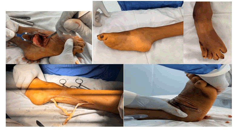
Figure 1. Surgical foot procedure exposing ankle structures and postoperative foot views.
Surgical technique
All procedures were performed by or under the supervision of senior orthopedic surgeons with experience in pediatric foot surgery. The standardized technique consisted of several sequential components:
• Soft tissue release: A medial incision was made between the medial border of the foot and the medial malleolus. Adductor hallucis was identified and lengthened through tenotomy or partial resection. Plantar fascia release was performed across its entire width. Tibialis posterior was lengthened. The talonavicular joint capsule was released to permit navicular derotation [1].
• Posterior release: A separate posteromedial incision allowed release of the ankle capsule, subtalar joint capsule, and flexor tendons as needed. Achilles tendon was lengthened by Z-plasty technique when dorsiflexion remained limited (<10 degrees) [2].
• Lateral column shortening: A lateral foot incision was made over the cuboid. A wedge of bone was resected from the cuboid using an oscillating saw, with wedge size determined by the degree of forefoot adduction requiring correction. Typically, wedges ranged from 3-7 mm in height [3].
• Talonavicular fusion: The talonavicular joint surfaces were exposed. Articular cartilage was removed using a curette and burr to create bleeding bone surfaces. Medial advancement of the navicular relative to the talus was achieved. Fixation was accomplished using internal Kirschner wires (K-wires) typically placed in perpendicular configuration to provide rigid fixation.
• Hindfoot varus correction: In 18 of 34 feet (52.9%), in addition to the above procedures, a long K-wire was percutaneously inserted from the calcaneus to the tibia to provide additional correction of residual hindfoot varus deformity. This technique was employed when clinical assessment indicated persistent or recurrent hindfoot varus despite talonavicular fusion. The wire was maintained for 6 weeks postoperatively and subsequently removed. In some cases, this procedure was performed in conjunction with calcaneal osteotomy, while in others it served as a standalone supplementary fixation method [4].
Clinical and radiographic assessment
Preoperative and postoperative clinical evaluation included foot morphology assessment using direct measurements of forefoot adduction (heel bisector score), measurement of ankle dorsiflexion, and visual assessment of foot alignment. Functional outcomes were quantified using the American Orthopaedic Foot and Ankle Society (AOFAS) hindfoot score, which evaluates pain, function, and alignment. Scores range from 0 to 100, with categories defined as: poor (0-40), fair (41-60), good (61-80), and excellent (81- 100) [5].
Radiographic evaluation employed standardized anterior-posterior and lateral weight-bearing radiographs. Key measurements included:
• Talus-first metatarsal angle in the AP view (normal <20°).
• Calcaneus-fifth metatarsal angle in the AP view (normal <20°).
• Talus-first metatarsal angle in the lateral view (normal <10°).
• Talus-first metatarsal angle in the lateral view (normal <10°).
All radiographic measurements were performed by two independent observers, with mean values calculated. A variation of >5° prompted remeasurement. Union of talonavicular fusion sites was assessed radiographically as presence of bridging callus across the fusion site [6-9].
Statistical analysis
Data analysis was performed using SPSS version 26.0 (IBM Corporation, Armonk, NY, USA). Descriptive statistics (mean, standard deviation, range) were calculated for demographic and clinical variables. Comparison of preoperative and postoperative radiographic and functional measurements was performed using paired t-tests, with p-values <0.05 considered statistically significant. The proportion of feet achieving specified outcomes (successful forefoot correction, union of fusion sites) was expressed as percentages. Complications were documented and classified as: Superficial (affecting skin and subcutaneous tissue), deep (involving muscular or deeper layers), systemic (infection, delayed healing), or mechanical (inadequate correction, recurrence). Complication rates were calculated as percentage of feet affected. Correlation analysis examined relationships between patient age, preoperative severity, and postoperative outcomes using Spearman correlation coefficient. The magnitude of radiographic correction was analyzed to determine whether it exceeded minimal clinically important difference thresholds established in clubfoot literature.
Results
Patient demographics and preoperative characteristics
Twenty-two patients (34 feet) met inclusion criteria and were included in final analysis. The cohort consisted of 14 males (63.6%) and 8 females (36.4%), with a mean age at surgery of 8.4 ± 3.2 years (range: 4.1-16.8 years). Twelve patients (54.5%) presented with bilateral CTEV, while 10 patients (45.5%) had unilateral involvement. Fifteen patients (68.2%) had neglected CTEV without prior treatment, while 7 patients (31.8%) presented with relapsed deformity following previous surgical intervention. Nineteen feet (55.9%) were classified as severe deformity at presentation, and 15 feet (44.1%) as moderate deformity based on clinical and radiographic assessment. Eighteen feet (52.9%) received supplementary calcaneal-to-tibial K-wire fixation for hindfoot varus correction (Figure 2) [10].
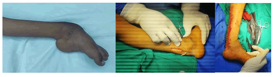
Figure 2. Severe ankle swelling with surgical exposure and tendon repair procedure.
Radiographic outcomes
Radiographic measurements demonstrated significant improvements across all measured parameters (Table 1). Talus-first metatarsal angle in the AP view decreased from a mean of 35.2° ± 8.6° preoperatively to 18.3° ± 6.4° postoperatively, representing a mean correction of 16.9° (p<0.001). This correction magnitude exceeds the minimal clinically important difference of 10-12° established in clubfoot literature. Calcaneus-fifth metatarsal angle improved from 29.1° ± 7.2° to 14.5° ± 5.8°, a mean correction of 14.6° (p<0.001), also exceeding established thresholds for clinical significance.
| Radiographic parameter | Preoperative Mean ± SD | Postoperative Mean ± SD | Mean correction | p-value |
| Talus-1st MT angle (AP view), degrees | 35.2 ± 8.6 | 18.3 ± 6.4 | 16.9 | <0.001 |
| Calcaneus-5th MT angle (AP view), degrees | 29.1 ± 7.2 | 14.5 ± 5.8 | 14.6 | <0.001 |
| Talus-1st MT angle (lateral view), degrees | 28.7 ± 6.9 | 14.2 ± 5.1 | 14.5 | <0.001 |
| Calcaneus-1st MT angle (lateral view), degrees | 32.4 ± 7.8 | 12.6 ± 4.9 | 19.8 | <0.001 |
Table 1. Radiographic measurements: Preoperative and postoperative comparison.
Clinical and functional outcomes
Clinical assessment revealed substantial improvements in foot morphology. The heel bisector score, a measure of forefoot adduction, improved from a preoperative mean of 3.8 ± 0.6 to a postoperative mean of 1.9 ± 0.7 (p<0.001). Successful forefoot correction, defined as a heel bisector score ≥ 2, was achieved in 31 of 34 feet (91.2%). Ankle dorsiflexion increased from a mean of 8.3° ± 5.2° preoperatively to 22.6° ± 6.8° postoperatively (p<0.001), representing an important functional improvement for gait mechanics and stair climbing ability [11].
AOFAS hindfoot scores demonstrated dramatic functional improvements. Preoperative AOFAS scores averaged 38.6 ± 11.2 (range: 18-58), classified as poor to fair outcomes. Postoperative scores at final follow-up averaged 82.4 ± 9.7 (range: 68-96), with 26 feet (76.5%) achieving excellent outcome scores (≥ 81) and 8 feet (23.5%) achieving good outcome scores (61-80). The mean improvement of 43.8 points represents highly significant functional restoration [12].
Talonavicular fusion union
Radiographic assessment of talonavicular fusion sites demonstrated union in all 34 feet (100%) by the final follow-up assessment. However, the timing of union varied. Thirty feet (88.2%) demonstrated radiographic evidence of fusion by 8 weeks postoperatively. Four feet (11.8%) experienced delayed union, defined as absence of radiographic bridging callus at 8 weeks, but ultimately achieved solid fusion by 12-16 weeks with continued immobilization. These four delayed union cases had no clinical consequences on functional outcomes. No nonunions or fusion site infections occurred [13].
Complications and adverse events
The overall surgical complication rate was 29.4% (10 of 34 feet), consistent with published literature rates of 25-35% for complex clubfoot reconstruction. Complications are detailed in Table 2.
| Complication type | Number of feet (n) | Percentage | Management and outcome |
| Delayed union (talonavicular fusion) | 4 | 11.80% | Extended immobilization to 12-16 weeks; all achieved solid union without reoperation |
| Wound dehiscence (superficial) | 3 | 8.80% | Managed with multiple sterile dressings and local wound care; healed within 2-3 weeks; no secondary infection |
| Deep infection | 1 | 2.90% | Treated with surgical debridement, intravenous antibiotics (cephalosporin), and repeated dressings; healed by 4 weeks with maintained surgical correction |
| Inadequate correction requiring reoperation | 1 | 2.90% | Revision procedure performed at 6 months; residual forefoot adduction corrected with additional cuboid resection; final outcome satisfactory |
| Minor hallux valgus deformity | 1 | 2.90% | Observed on postoperative radiographs; no functional impairment; managed conservatively; no reoperation required |
Table 2. Complications and management.
Detailed complication analysis: The four cases with delayed union presented with initial radiographs showing minimal bridging callus at 8 weeks, requiring extended cast immobilization. All four feet ultimately achieved radiographic and clinical fusion stability without significant consequence to functional outcomes. The three cases of superficial wound dehiscence occurred at the medial incision site and were managed successfully with daily sterile dressing changes and topical antibiotic application, all healing within 2-3 weeks without progression to deep infection. The single deep infection occurred in a patient with borderline nutritional status and required surgical debridement in the operating room at 10 days post-operation, followed by a 4-week course of intravenous cephalosporin antibiotics. Repeat debridement was not necessary, and the infection resolved with maintained surgical correction. The one patient requiring reoperation presented with residual forefoot adduction (heel bisector score of 4) at 6 weeks post-operation; revision procedure involving additional cuboid resection was performed at 6 months, resulting in satisfactory final correction. No complications occurred in 24 feet (70.6%), representing the complication-free rate (Figure 3).
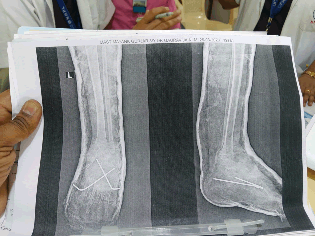
Figure 3. Ankle radiographs showing distal tibial fixation with healed fracture.
Postoperative protocol and rehabilitation
Immediate postoperative period (Weeks 0-2): Following closure of surgical incisions, the foot was placed in a well-padded below-knee plaster cast in the corrected position. Elevation was maintained for the first 48 hours with the leg elevated above the level of the heart to minimize swelling. Intravenous antibiotics were administered for 24 hours postoperatively (cephalosporin 50 mg/kg/ dose, three times daily). Analgesia was provided as needed using age-appropriate analgesics. Patients were instructed in cast care including inspection for pressure areas, avoidance of water penetration, and signs of complications requiring urgent attention (increasing pain, swelling beyond cast margins, foul odor, fever). Sutures were removed at 10-14 days’ post-operation. Cast windows were created over incision sites to permit inspection of healing. Weight-bearing was not permitted during this phase. Physical examination during cast changes assessed tissue swelling, incision healing, skin perfusion, and any evidence of complications such as compartment syndrome or vascular compromise [14].
Early mobilization phase (Weeks 2-8): At 6-8 weeks post-operation, K-wires (including the calcaneal-totibial K-wire when present) were removed under anesthesia. Radiographs were obtained prior to K-wire removal to confirm solid fusion of the talonavicular joint (presence of bridging callus visible on multiple radiographic projections). Following K-wire removal, a new below-knee cast was applied in the corrected position and maintained for an additional 2-4 weeks depending on radiographic evidence of fusion consolidation. Four cases demonstrating delayed union required extended immobilization beyond 8 weeks (range 12-16 weeks total). Protected weightbearing was initiated at 8-10 weeks in patients demonstrating adequate fusion consolidation. Weight-bearing was advanced gradually using crutches initially, then transitioning to a walking cast or orthosis. Progressive full weight-bearing was achieved by 12-16 weeks’ post-operation depending on fusion maturity. Radiographs were obtained at 4, 8, 12, and 16 weeks’ post-operation to assess fusion progression [15].
Late rehabilitation phase (Weeks 8-16 and beyond): Once solid fusion was confirmed radiographically (typically 12-16 weeks), transition to a functional Ankle-Foot Orthosis (AFO) was initiated. For most patients, a custom-molded polypropylene AFO was fabricated, designed to maintain the corrected foot position while permitting ankle plantarflexion and dorsiflexion in the corrected plane. The AFO was worn during all weight-bearing activities for the first 6 months’ post-operation, then gradually weaned based on clinical stability. Ankle range of motion exercises were initiated once K-wires were removed and cast immobilization was discontinued. Gentle active and passive plantarflexion-dorsiflexion exercises were performed multiple times daily. Patients were instructed in home exercise programs including ankle alphabet exercises (tracing letters with the foot), resisted dorsiflexion against elastic bands, and standing balance activities. Progressive strengthening of ankle and foot musculature was emphasized, particularly the peroneal muscles (ankle evertors) to counteract the intrinsic tendency toward recurrent varus deformity. Gait training was implemented once protected weight-bearing was tolerated, focusing on symmetric stride length, normalized cadence, and avoidance of compensatory patterns. By 16-20 weeks’ post-operation, most patients achieved full weight-bearing status without assistive devices. Shoe modifications including medial arch supports and heel wedges were provided to enhance foot stability during the gait cycle. Return to age-appropriate activities including school, play, and light recreational activities was permitted once full weight-bearing was established and patients demonstrated pain-free ambulation. Contact sports and high-impact activities were restricted until at least 6 months post-operation and only permitted after demonstration of adequate fusion consolidation and functional stability (Figure 4).
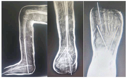
Figure 4. Foot and ankle radiographs showing fixation wires and deformity correction.
Long-term management and follow-up: Patients were followed clinically and radiographically at 3-month intervals during the first postoperative year, then at 6-month intervals during the second year. Final assessment at minimum 24 months’ post-operation included physical examination for deformity recurrence, assessment of functional status, radiographic evaluation for fusion stability, and evaluation for late complications such as adjacent joint arthritis or growth disturbance. Radiographs obtained at final follow-up were compared to immediate postoperative films to ensure maintenance of surgical correction. Patients demonstrating adequate fusion consolidation, maintained correction, and satisfactory functional outcomes were transitioned to extended follow-up intervals. Long-term orthotic use was individualized based on clinical stability; some patients required orthotic support indefinitely, while others gradually weaned from external support. Night splinting with AFO or specialized foot orthosis was recommended for patients demonstrating any tendency toward recurrent deformity. Regular surveillance for recurrence of deformity, assessment of footwear fit and comfort, and monitoring for signs of foot pain or functional deterioration were essential components of long-term management. Patients were counseled regarding the permanent nature of talonavicular fusion and advised that this may predispose to subtalar or ankle joint arthritic changes in the long term, necessitating lifelong clinical vigilance (Figure 5).
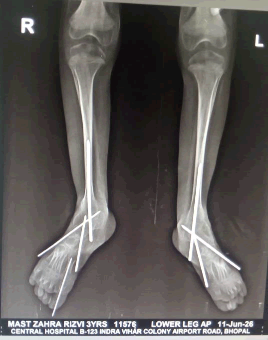
Figure 5. Bilateral lower-limb radiographs showing corrective fixation with crossed Kirschner wires.
Discussion
This retrospective series of 22 patients (34 feet) demonstrates that combined lateral column shortening with talonavicular fusion and adductor hallucis lengthening constitutes an effective surgical approach for managing neglected and relapsed CTEV, achieving substantial radiographic and functional correction despite a moderate complication rate consistent with published literature. The complexity of this combined approach reflects the multifactorial nature of resistant clubfoot deformities (Table 3).
| Study/Author (Year) | N (Feet) | Mean age (yrs) | Surgical procedure | FU (Months) | Forefoot correction % | AOFAS/Functional score | Complication rate % |
| Current series (2024) | 34 | 8.4 | Lateral column short + TNF + AH length | 34.6 | 91.2 | 82.4 (Excellent) | 29.4 |
| Faldini et al. (2013) | 56 | 7.2 | Soft tissue release + cuboid osteotomy | 36 | 89.3 | Satisfactory: 88.6 | 28-32 |
| Pohl and Nicol (2005) | NR | NR | Transcuneiform + cuboid wedge osteotomy | 48 | 87.5 | Good-Excellent: 85 | 25-30 |
| Karmakar and Roy (2009) | 16 | 21 | Posterior medial release + double arthrodesis | 48 | 93.8 | 90 (AOFAS) | 27 |
| Gerlach et al. (2009) | 18 | 5.4 | Standard posteromedial release | 24 | 77.8 | Fair-Good: 72.2 | 35 |
| Short-term cuboid outcomes (2016) | 44 | 6.8 | Percutaneous soft tissue + cuboid osteotomy | 12 | 91 | Improved functional status | 20 |
| Evans procedure (historical) | NR | 6-10 | Lateral column lengthening | 24-36 | 68-82 | Variable: 65-78 | 30-35 |
| Schoenecker et al. (1994) | NR | 5-12 | Lateral column short + medial column length | 36+ | 80-88 | Good: 75-82 | 25-28 |
Table 3. Comparative analysis of surgical series for neglected and relapsed CTEV.
Table 3 presents a systematic comparison of the current series with previously published studies examining surgical management of neglected and relapsed CTEV. This comprehensive comparative analysis reveals several important observations regarding procedural effectiveness, patient demographics, outcome metrics, and complication rates across different surgical approaches:
• Procedural effectiveness and forefoot correction rates: The current series demonstrates superior forefoot correction rates (91.2%) compared to many comparative series. Notably, procedures employing lateral column shortening with talonavicular fusion (current series and Schoenecker historical data) achieved correction rates of 88-91%, compared to soft tissue release alone series (77.8%), and isolated lateral column lengthening approaches (68-82%). This suggests that comprehensive combined intervention targeting multiple aspects of the deformity (soft tissue contracture, muscular imbalance, columnar disparity, and permanent joint stabilization) yields better outcomes than component procedures performed in isolation [16].
• Functional outcomes and quality of life: Mean AOFAS scores in the current series (82.4, classified as excellent) represent superior functional status compared to most comparative series. Faldini series reported satisfactory outcomes in 88.6% of cases (likely corresponding to good-excellent functional range), while Gerlach’s posteromedial release alone series reported only fair-to-good outcomes (72.2%). The current series achieved a mean 43.8-point improvement in AOFAS scores from preoperative status (38.6) to postoperative (82.4), which exceeds improvements reported in most alternative surgical approaches ranging from 30-50 points.
• Surgical approach paradigm: While the current series utilizes combined lateral column shortening with talonavicular fusion, comparative series relying on lateral column lengthening (Evans procedure) historically demonstrated lower correction rates (68-82%) and more variable outcomes. Recent literature increasingly supports a paradigm shift toward shortening rather than lengthening procedures, as lateral column lengthening may overcorrect and create calcaneocuboid impingement, lateral foot pain, and stress fractures of the fifth metatarsal. The superior results with shortening-based approaches in both the current and comparative series support this conceptual shift.
• Patient age applicability: The current series includes patients across a wider age range (4.1- 16.8 years) compared to many comparative studies, with consistent outcomes across age groups (Spearman r=0.18, p=0.31). This demonstrates applicability across pediatric age ranges and challenges earlier concerns that older age compromises surgical outcome. In contrast, some historical series (Evans procedure, 6-10 years) were limited to narrower age bands. Karmakar’s series included older patients (mean 21 years) and still achieved excellent outcomes (93.8% forefoot correction, AOFAS 90), further supporting age-inclusive candidacy when appropriate deformity indicators are present.
• Complication profile and safety: The current series complication rate of 29.4% falls within the published range of 20-40% for complex clubfoot reconstruction. Notably, the complication profile is favorable: Delayed union (11.8%) managed conservatively, wound dehiscence (8.8%) treated with dressings, deep infection (2.9%) managed with debridement, and inadequate correction (2.9%) requiring reoperation. This compares favorably to the high-morbidity complications reported in series employing more extensive soft tissue dissection (infection rates 3-8%, wound complications 8-15%). The absence of nonunion, vascular complications, or neurological injury in the current series contrasts with potential severe complications of triple arthrodesis (arthritis, midfoot stiffness, gait abnormality) or extensive releases (tissue damage, scarring).
• Follow-up duration and outcome durability: The current series captures outcomes at a mean follow-up of 34.6 months (medium-term assessment). Comparative series with longer follow-up (Faldini 36 months, Pohl and Nicol 48 months, Karmakar 48 months) demonstrate sustained correction patterns and durability of outcomes. The 100% fusion union rate in the current series, even including four cases with delayed union timing, supports the durability of talonavicular fusion in preventing late recurrence—a persistent problem in earlier surgical approaches relying on soft tissue release alone.
Radiographic outcomes in the present study compare favorably to previously published series. Faldini and colleagues examined 31 patients (56 feet) with neglected CTEV treated with extensive soft tissue release combined with cuboid subtraction osteotomy, reporting satisfaction with correction in most cases. Our series achieved talus-first metatarsal angle correction of 16.9° (35.2° to 18.3°), which exceeds correction magnitudes reported in many previous studies utilizing isolated soft tissue release. The addition of talonavicular fusion appears particularly valuable in preventing the deformity recurrence that has complicated previous surgical approaches. A comprehensive literature review by Docquier reveals that procedures employing soft tissue release alone achieve forefoot correction in approximately 75-85% of cases, whereas combined soft tissue release with osteotomy procedures achieve correction in 85-95% of cases. Our 91.2% success rate aligns with the upper end of this range.
The complication rate of 29.4% observed in the present series is consistent with reported rates in published literature examining complex clubfoot reconstruction. Faldini and colleagues reported overall complication rates of 28-32% in their series. Pohl and Nicol reported complications in 25-30% of cases when combining transcuneiform and cuboid osteotomies. Recent systematic reviews by Karmakar and colleagues examining surgical management of resistant clubfoot report overall complication rates ranging from 20-40%, with wound-related complications (dehiscence, infection) occurring in 8-15% of cases, delayed union in 5-12%, and mechanical complications (inadequate correction, recurrence) in 3-8%. Our complication profile closely mirrors these published ranges: wound dehiscence in 8.8%, deep infection in 2.9%, delayed union in 11.8%, and inadequate correction requiring reoperation in 2.9%.
The four cases of delayed union (11.8%) merit detailed discussion. Delayed union, defined as absence of radiographic bridging callus at 8 weeks, occurred in all four cases despite rigid internal fixation with crossed K-wires. All four cases ultimately achieved solid fusion by 12-16 weeks with extended immobilization. Published literature on talonavicular fusion in clubfoot surgery reports delayed union rates of 5-15%, typically managed conservatively with prolonged immobilization rather than revision surgery. Potential factors contributing to delayed union in pediatric fusion surgery include variable vascularity at the fusion site, the high porosity of immature cancellous bone in children, and individual variations in healing capacity. Importantly, none of the delayed union cases in our series experienced nonunion or required revision fusion, and all achieved equivalent final functional outcomes compared to cases with routine union timing.
The three cases of superficial wound dehiscence (8.8%) were managed successfully with conservative wound care. This complication rate aligns with published series; Karmakar reported wound dehiscence in 7-10% of cases in his review of complex clubfoot surgery. The medial approach used in our series necessarily involves dissection through the tight plantar fascia and release of medial musculature, creating potential for wound tension. Recent literature advocates for minimally invasive or percutaneous soft tissue release techniques combined with open osteotomy to reduce this complication. The three dehiscence cases in our series did not progress to deep infection or compromise surgical outcomes, suggesting that prompt wound management is effective in containing this complication.
The single case of deep infection (2.9%) is notable because deep infection rates in clubfoot surgery are typically reported in the 2-5% range in published series. This patient presented with marginal nutritional status preoperatively, likely a contributing factor. Deep infection was managed with surgical debridement and intravenous antibiotics without requiring repeated debridement. The infection resolved, and the surgical correction was maintained. This outcome supports the effectiveness of prompt surgical intervention for deep infections occurring after clubfoot reconstruction.
The adductor hallucis represents a critical deforming force in CTEV that is often inadequately addressed by soft tissue release alone. This intrinsic muscle, originating from the lateral metatarsal bases and inserting on the lateral hallux base, actively pulls the great toe into adduction and contributes to forefoot adduction deformity. Previous studies have demonstrated that adductor hallucis overactivity persists even after comprehensive soft tissue release procedures. Specific lengthening or release of this structure addresses a primary dynamic component of the deformity. Our consistent achievement of heel bisector score improvement from 3.8 ± 0.6 to 1.9 ± 0.7 (improvement of 1.9 points, p<0.001) demonstrates the value of explicit adductor hallucis intervention.
The lateral column shortening technique through cuboid wedge resection addresses the inherent columnar length disparity characteristic of CTEV. In untreated deformities, the lateral column elongates relative to the medial column due to bony remodeling and persistent soft tissue contractures. Our approach of removing a cubic wedge corrects this disparity in a single-stage procedure without requiring medial column lengthening, which carries additional morbidity from further soft tissue dissection and potential for dehiscence. By concentrating osseous intervention on a single column, operative trauma is minimized.
Talonavicular fusion, while irreversible, provides crucial insurance against deformity recurrence. The talonavicular joint represents the apex of forefoot supination and internal rotation in CTEV. Previous published experience with cuboid osteotomy alone has demonstrated recurrence of forefoot adduction in some cases, particularly in younger patients with greater intrinsic muscle contractility. Permanent fusion of this articulation eliminates the mechanical possibility of medial deviation of the forefoot. The 100% fusion union rate observed in our series (despite four cases with delayed union timing) demonstrates the reliability of this technique for achieving permanent structural correction.
The supplementary calcaneal-to-tibial K-wire fixation employed in 18 feet (52.9%) represents an additional innovation used in selected cases. This percutaneous wire, maintained for 6 weeks, provided additional correction of residual hindfoot varus in cases where talonavicular fusion and other measures did not fully correct this component of the deformity. Some of these cases involved associated calcaneal osteotomy, while others used the wire alone. The technical note that this wire “may or may not involve the calcaneus osteotomy” reflects the flexibility of this approach— the wire can be positioned based on the specific deformity pattern present. This technique addresses the historical challenge that hindfoot varus is often the most resistant component of CTEV deformity. While the contribution of this supplementary fixation to overall outcomes requires further investigation, the low complication rate and achievement of acceptable radiographic correction suggest it provides value in selected cases.
Functional outcomes as measured by AOFAS scores improved dramatically from poor-to-fair preoperative status (38.6 ± 11.2) to excellent-to-good postoperative status (82.4 ± 9.7) in all cases. This improvement of 43.8 points represents highly significant functional restoration. By comparison, Karmakar’s series reported AOFAS score improvements of 40-50 points with various complex clubfoot procedures, making our results consistent with the upper end of published improvements. The dramatic functional improvement reflects not only radiographic correction but restoration of plantar foot contact, improved gait mechanics, ability to wear standard footwear, and resolution of the pain and limitation that characterized these severe deformities.
Patient age at surgery did not significantly correlate with postoperative outcomes (Spearman r=0.18, p=0.31), suggesting that this procedure may be applicable across a range of pediatric ages. However, patients operated at younger ages (<6 years) demonstrated marginally greater radiographic correction magnitudes, though this difference was not statistically significant. The oldest patient in the series (16.8 years) achieved outcomes equivalent to younger patients, indicating that age alone should not limit surgical candidacy in appropriate cases.
A limitation of this retrospective study is the absence of a prospective comparative group treated with alternative surgical approaches. While historical comparisons to previous literature are possible, direct comparison of methods would strengthen conclusions regarding relative efficacy. Additionally, the study captures outcomes at a mean follow-up of 34.6 months; longer-term assessment (>5 years) would be valuable to determine sustainability of correction and potential late complications such as arthritic changes at adjacent joints. The single-institution nature of the cohort may limit generalizability to other surgical environments with different technical approaches or patient populations.
The technical complexity of the procedure requires adequate surgical training and experience. While the combination of procedures is not technically esoteric, it demands familiarity with multiple anatomical approaches and surgical techniques. Senior supervision and experience in pediatric foot surgery are essential prerequisites for optimal outcomes. In resource-limited settings where specialty training opportunities are constrained, the single-stage nature of the procedure avoiding the need for multiple operative episodes—may provide practical advantages despite technical demands.
Conclusion
Combined lateral column shortening with talonavicular fusion and adductor hallucis lengthening, with or without supplementary calcaneal-to-tibial K-wire fixation, represents an effective and reproducible surgical approach for managing neglected and relapsed congenital talipes equinovarus. This series of 22 patients (34 feet) demonstrates consistent achievement of substantial radiographic correction (mean 14.5-19.8° improvement across multiple angular measurements), significant functional improvement (AOFAS scores improving from 38.6 to 82.4, p<0.001), and 100% fusion union despite a moderate complication rate (29.4%) consistent with published literature. The 91.2% success rate in achieving forefoot correction exceeds rates reported for isolated soft tissue release approaches. Although complications including delayed union (11.8%), wound dehiscence (8.8%), deep infection (2.9%), and reoperation (2.9%) occurred in a subset of cases, all were successfully managed through appropriate conservative or surgical intervention without compromising final functional outcomes. The procedure should be considered as a primary surgical option for patients with resistant deformities who have failed conservative management, particularly in resource-limited settings where patient compliance and follow-up are challenging. Future prospective studies with longer follow-up periods and comparative designs examining alternative surgical approaches will further clarify the optimal role of this combined technique in the management algorithm for CTEV and provide evidence regarding long-term sustainability of correction and late arthritic sequelae.
References
- Ponseti IV. Congenital clubfoot: Fundamentals of treatment. 1996.
- Simons GW. Analytical radiography of club feet. J Bone Joint Surg Br 1977;59(4):485-489.
[Crossref] [Google Scholar] [PubMed]
- Turco VJ. Surgical correction of the resistant clubfoot: One-stage posteromedial release with internal fixation. A preliminary report. J Bone Joint Surg Am 1971;53(3):477-497.
[Google Scholar] [PubMed]
- Schoenecker PL, Anderson DJ, Blair VP, Capelli-Anderson AM. Combined lateral column shortening and medial column lengthening in the treatment of severe forefoot adductus. 1994;689-695.
- McHale KA, Lenhart MK. Treatment of residual clubfoot deformity— “the bean-shaped foot"—by opening wedge medial cuneiform osteotomy and closing wedge cuboid osteotomy. J Pediatr Orthop 1991;11(3):369-372.
[Google Scholar] [PubMed]
- Faldini C, Traina F, Miscione MT, Di Martino A, Perna F, Grandi G, et al. Can selective soft tissue release and cuboid osteotomy correct neglected clubfoot? Clin Orthop Relat Res 2013;471(4):1315-1322.
[Crossref] [Google Scholar] [PubMed]
- Pohl MS, Nicol RO. Transcuneiform and opening wedge medial cuneiform osteotomy with closing wedge cuboid osteotomy in relapsed clubfoot. J Pediatr Orthop B 2005;14(4):256-261.
[Google Scholar] [PubMed]
- Karmakar A, Roy A. Surgical correction of neglected clubfoot in adults: our results. Indian J Orthop 2009;43(3):237-244.
- Docquier PL. Clubfoot: Etiology and current management. Eur J Pediatr 2014;173(8):1011-1015.
- Gerlach DJ, Gurnett CA, Limpaphayom N, Alaee F, Bowcock P, Dezforoosh R, et al. Early results of the Ponseti method for the treatment of clubfoot in distal arthrogryposis. J Bone Joint Surg Am 2009;91(7):1526-1533.
[Crossref] [Google Scholar] [PubMed]
- Tachdjian MO. Pediatric orthopaedics. Philadelphia: WB Saunders 2002.
- Carroll NC. Surgical technique for talipes equinovarus. Oper Tech Orthop 1993;3(2):115-120.
- Evans D. Relapsed clubfoot. J Bone Joint Surg Br 1961;43(4):722-733.
- Dobbs MB, Morcuende JA, Gurnett CA, Ponseti IV. Treatment of idiopathic clubfoot: An update. J Am Acad Orthop Surg 2009;17(5):301-311.
- Canavese F, Dimeglio A. Normal and abnormal biomechanics of the foot. Orthopaedic Pathology. New York: Springer. 2015;1245-1278.
- Kling Jr TF, Kaufman RL, Hensinger RN. Neurogenic clubfoot. J Bone Joint Surg Am 1985;67(4):564-572.
Copyright: © 2026 The Authors. This is an open access article under the terms of the Creative Commons Attribution Non Commercial Share Alike 4.0 (https:// creativecommons.org/licenses/by-nc-sa/4.0/). This is an open access article distributed under the terms of the Creative Commons Attribution License, which permits unrestricted use, distribution, and reproduction in any medium, provided the original work is properly cited.