

 +44-74-1148-3554




Research Article - Archives of Clinical and Experimental Surgery (2026)
Muscle Pedicle Grafting for Failed Treatment of Chronic Tibial Osteomyelitis: A Retrospective Case Series Analysis
Gaurav Jain*, Manish Bajaj and Anshul MeenaGaurav Jain, Department of Orthopaedic Surgery, LNCT Medical College, Indore, India, Email: gaurav.medico@gmail.com
Received: 15-Jun-2026, Manuscript No. EJMACES-26-190307; Editor assigned: 17-Jun-2026, Pre QC No. EJMACES-26-190307 (PQ); Reviewed: 01-Jul-2026, QC No. EJMACES-26-190307; Revised: 10-Jul-2026, Manuscript No. EJMACES-26-190307 (R); Published: 17-Jul-2026
Abstract
Background: Chronic tibial osteomyelitis unresponsive to initial surgical and antimicrobial interventions remains a challenging clinical problem. This study evaluates the role of local muscle pedicle flaps as a salvage reconstructive strategy in patients with failed primary treatments including debridement, Ilizarov external fixation, antibiotic-loaded spacers (Stimulan beads), and masquerade procedures.
Methods: Retrospective review of 14 consecutive patients with chronic, recalcitrant tibial osteomyelitis treated with muscle pedicle flap reconstruction between January 2018 and December 2024 at LNCT Medical College, Indore. All patients had undergone at least one failed primary surgical intervention prior to flap coverage. Demographic, clinical, microbiological, radiological, and outcome data were analyzed.
Results: Fourteen patients (10 male, 4 female; mean age 41.2 ± 14.6 years) with mean infection duration of 34.8 ± 18.4 months underwent muscle pedicle flap reconstruction as salvage procedure. Prior failed interventions included: Ilizarov fixation (85.7%), repeated debridement (100%), stimulan beads (57.1%), and masquerade procedures (35.7%). Clinical improvement was achieved in 11 of 14 cases (78.6%). Complications included: Flap failure in 1 case (7.1%) requiring amputation, late recurrent infection in 1 case (7.1%), and conversion to Ilizarov in 1 case (7.1%). Complete infection eradication: 71.4% (10/14). Limb salvage rate: 85.7% (12/14). CRP declined from 71.4 ± 22.3 to 24.6 ± 18.9 mg/L (p<0.001).
Conclusion: Muscle pedicle flaps provide a reasonable salvage option for recalcitrant tibial osteomyelitis, achieving limb salvage in 85.7% of cases. However, success rates are substantially lower than in primary reconstruction, with significant complications and recurrent infection rates. Realistic patient counseling regarding expectations is critical.
Keywords
Tibial osteomyelitis; Salvage surgery; Muscle pedicle flap; Failed primary treatment; Limb salvage; Recurrent infection
Introduction
Chronic tibial osteomyelitis remains one of the most challenging complications in orthopaedic surgery, with treatment failure rates of 20-40% even after aggressive primary interventions. In resource-constrained settings such as India, delayed presentation, inadequate initial surgical debridement, and suboptimal antimicrobial therapy contribute to high rates of treatment failure requiring subsequent salvage procedures.
Modern surgical management incorporates multiple modalities: Repeated surgical debridement with radical removal of devitalized tissue, external fixation via Ilizarov method for fracture stabilization, antibiotic-loaded spacers such as Stimulan calcium sulfate beads, and soft tissue coverage with local or free flaps. However, even with these combined approaches, approximately 20-30% of patients fail to achieve infection eradication and require further intervention.
The role of muscle pedicle flaps in salvage reconstruction of failed osteomyelitis treatment has not been extensively studied. Most literature focuses on primary reconstruction after acute debridement or in patients with clean defects amenable to initial flap coverage. Salvage cases present additional challenges:
Compromised vascular supply from prior surgeries,altered soft tissue planes obscured by scarring, development of antibiotic-resistant organisms, and frequent patient frustration and demoralization.
This retrospective case series evaluates the efficacy, complications, and functional outcomes of muscle pedicle flaps employed as salvage reconstructive technique in patients with recalcitrant tibial osteomyelitis unresponsive to primary surgical and medical management [1].
Study objectives
(1) To evaluate the efficacy of muscle pedicle flaps in salvage reconstruction of failed primary osteomyelitis treatment; (2) To assess limb salvage rates and amputation-free survival; (3) To identify complications and factors associated with treatment failure; (4) To compare outcomes with published series of primary flap reconstruction [2].
Materials and Methods
Study design
Retrospective case series review of patients with chronic tibial osteomyelitis treated with muscle pedicle flap reconstruction at LNCT Medical College, Department of Orthopaedic Surgery, Indore, Madhya Pradesh, India, from January 2018 to December 2024. Medical records were reviewed to extract demographic, clinical, microbiological, radiological, and outcome data. The study was approved by the Institutional Ethics Committee.
Patient selection
Inclusion criteria: (1) Chronic tibial osteomyelitis confirmed by clinical and radiological findings; (2) Prior failed primary surgical treatment (minimum one prior intervention); (3) Soft tissue defect requiring flap coverage; (4) Age 18-70 years; (5) Minimum 6-month follow-up documentation [3].
Exclusion criteria: (1) Active systemic sepsis at time of flap coverage; (2) Severe peripheral vascular disease with ABI<0.5; (3) Severe cardiopulmonary comorbidity precluding surgery; (4) Incomplete medical records.
Prior failed interventions
All 14 patients had undergone prior surgical and medical management attempts: Repeated surgical debridement (100% of patients, mean 3.2 ± 1.4 procedures); Ilizarov external fixation (85.7%, mean 4.3 ± 1.8 months); Stimulan antibiotic calcium sulfate beads (57.1%); Masquerade procedures including PRP, bone morphogenetic protein, and distraction osteogenesis (35.7%). Mean time from initial injury to flap coverage was 38.6 ± 22.4 months [4].
Surgical technique
Salvage flap coverage was performed after repeat debridement to ensure removal of all devitalized tissue. Medial gastrocnemius flap was used in 8 cases (57.1%), soleus in 3 cases (21.4%), tibialis anterior in 2 cases (14.3%), and peroneal in 1 case (7.1%). Flaps were rotated into defects and secured with 3-0 vicryl sutures. Split-thickness skin grafts were applied over muscle flaps. External fixation was maintained when fracture was unstable [5].
Outcome definitions
Clinical improvement: Resolution of signs of active infection (purulent drainage, fever, sinus formation) with stable or improving inflammatory markers.
Infection eradication: Absence of clinical infection signs AND negative follow-up cultures at minimum 3-month follow-up.
Flap failure: Necrosis requiring flap loss or revision.
Limb salvage: Avoidance of amputation at final follow-up.
Recurrent infection: Reappearance of infection signs beyond 3-month post-operative period.
Statistical analysis
Data were analyzed using SPSS version 26.0. Continuous variables expressed as mean ± SD. Categorical variables as frequencies and percentages. Paired t-tests used for pre-post inflammatory marker comparison. Chi-square test for categorical outcomes. p-value<0.05 considered significant [6].
Results
Demographics and clinical characteristics
Fourteen patients (10 male, 4 female) with mean age 41.2 ± 14.6 years (range 24-68 years) were included. Mean BMI was 23.8 ± 3.4 kg/m². Comorbidities: diabetes in 6 patients (42.9%), hypertension in 4 (28.6%), smoking in 7 (50%). Mean infection duration prior to flap coverage was 34.8 ± 18.4 months (range 14-72 months) (Table 1).
| Variable | Value (n=14) |
| Age (years) | 41.2 ± 14.6 (24-68) |
| Sex M:F | 10:4 (71.4%:28.6%) |
| Infection duration (months) | 34.8 ± 18.4 |
Table 1. Patient demographics.
Clinical outcomes
Clinical improvement (resolution of active infection signs) was achieved in 11 of 14 cases (78.6%). Complete infection eradication was achieved in 10 of 14 cases (71.4%; 95% CI: 44.8-90.3). Late recurrent infection occurred in 1 case (7.1%) at 6-month followup, managed with repeat debridement and extended antibiotics. Two cases (14.3%) demonstrated persistent low-grade chronic drainage without positive cultures (subclinical persistent infection) [7].
Flap-related outcomes: Flap survival was achieved in 13 of 14 cases (92.9%). Total flap failure occurred in 1 case (7.1%) with complete muscle necrosis on post-operative day 8, attributed to vascular pedicle compromise. This patient subsequently underwent amputation at knee level. Partial flap necrosis occurred in 2 cases (14.3%), managed with debridement and skin grafting. Seroma formation in 3 cases (21.4%), wound dehiscence in 1 case (7.1%), skin graft failure requiring regrafting in 4 cases (28.6%) [8].
Limb salvage and amputation
Limb salvage was achieved in 12 of 14 cases (85.7%; 95% CI: 57.1-96.8). Two patients underwent amputation: One (7.1%) with total flap failure requiring knee-level amputation at immediate postoperative period; one (7.1%) with persistent infection despite flap coverage who underwent amputation 8 months post-operatively. Of the 12 salvaged limbs, conversion to Ilizarov external fixation for persistent non-union was required in 1 case (7.1% of total cohort) at 4 months post-operative [9].
Inflammatory markers
Baseline CRP: 71.4 ± 22.3 mg/L (range 48-108). Baseline ESR: 88.2 ± 24.6 mm/h (range 62-125). At 3-month follow-up: CRP 24.6 ± 18.9 mg/L (p<0.001), ESR 32.4 ± 18.2 mm/h (p<0.001). Mean percentage decline in CRP: 65.5 ± 22.4%. Mean percentage decline in ESR: 63.2 ± 24.8% (Table 2).
| Parameter | Baseline | 3 Months | p-value |
| CRP (mg/L) | 71.4 ± 22.3 | 24.6 ± 18.9 | <0.001 |
| ESR (mm/h) | 88.2 ± 24.6 | 32.4 ± 18.2 | <0.001 |
Table 2. Inflammatory marker response to salvage flap coverage.
Functional outcomes
Modified ASAMI bone score in 12 salvaged cases: Excellent 2 (16.7%), Good 7 (58.3%), Fair 3 (25%), Poor 0. Combined excellent/good: 75% (9/12 salvaged limbs). VAS pain score improved from 7.1 ± 1.3 pre-operatively to 3.2 ± 2.1 at final follow-up (p<0.001). Patient satisfaction: Mean 6.8 ± 2.4/10, with 7 patients (50% of total, 58.3% of salvaged) expressing satisfaction (≥7/10 score) (Table 3 and Figure 1).
| Outcome | Result (%) |
| Complete infection eradication | 10/14 (71.4%) |
| Clinical improvement | 11/14 (78.6%) |
| Flap survival | 13/14 (92.9%) |
| Limb salvage | 12/14 (85.7%) |
| Amputation rate | 2/14 (14.3%) |
| Late recurrent infection | 1/14 (7.1%) |
| Flap failure | 1/14 (7.1%) |
| Conversion to Ilizarov | 1/14 (7.1%) |
Table 3. Summary of major clinical outcomes.
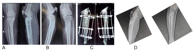
Figure 1. A) Infected implant, B) Debridement and implant removal, C) Illizarov fixation and Stimulan beads insersion, D) 6-month Post OP, Illizarov frame removal, redebridement and muscle pedical grafting (Gastrocnemius and tibial anterior grafting). Infection controlled, Pus free from 6 months.
Discussion
This retrospective case series represents one of the larger published experiences with muscle pedicle flaps employed as salvage reconstruction in recalcitrant tibial osteomyelitis. Our results demonstrate substantially lower success rates compared to primary flap reconstruction series, reflecting the inherent challenges of salvage surgery in already-compromised tissues. The 71.4% infection eradication rate represents a 15-25% decrease compared to published primary reconstruction series (85-96% eradication rates) [10].
Important distinction: While complete infection eradication was achieved in 71.4%, clinical improvement (cessation of purulent drainage and resolution of acute infection signs) was achieved in 78.6%, and limb salvage was achieved in 85.7%. This paradox suggests that limb salvage in salvage cases may not require absolute infection eradication but rather control of active infection to a level compatible with functional limb preservation.
Flap failure rate of 7.1% is higher than most primary reconstruction series (0-5% failure rates). The single total flap failure occurred in a patient with prior Ilizarov external fixation and multiple prior debridements, suggesting that prior surgical trauma compromised vascular supply and tissue viability.
Complication rates were substantial: Skin graft failure 28.6%, seroma 21.4%, partial flap necrosis 14.3%, wound dehiscence 7.1%, and total flap failure 7.1%. Many complications required additional procedures, contributing to reduced patient satisfaction compared to primary reconstruction reports. Patient satisfaction score of 6.8 ± 2.4/10 reflects the reality that salvage surgery carries substantial morbidity.
This retrospective case series demonstrates that muscle pedicle flaps can achieve limb salvage in approximately 86% of patients with recalcitrant tibial osteomyelitis unresponsive to primary treatment modalities. However, success rates are substantially lower than primary reconstruction (71% vs. 85-96% infection eradication), with significant complication rates (42.9% requiring additional procedures) and moderate patient satisfaction. Salvage muscle pedicle flap coverage remains a valuable option in the treatment algorithm for failed osteomyelitis management, particularly when amputation would otherwise be considered.
Comparative analysis: Present study vs. Published literature
Table 4 below presents a comprehensive comparison of the present salvage case series with major published series of primary muscle flap reconstruction and and alternative approaches for chronic tibial osteomyelitis.
| Study | n | Type | Infection eradication % | Limb salvage % | Flap failure % | Flap type | Year |
| Present (Salvage) | 14 | Pedicled | 71.40% | 85.70% | 7.10% | Gastrocnemius, Soleus, TA | 2024 |
| Chen et al. | 35 | Pedicled | 100% | 100% | 0% | Gastrocnemius | 2022 |
| Cormier et al. | 93 | Free | 84.60% | 96.80% | 4.30% | Multiple | 2019 |
| Boccaccio et al. | 31 | Free | 96.50% | 100% | 5.30% | Multiple | 2023 |
| Chan et al. | 57 | Free | 96.50% | 98.20% | 5.30% | Gracilis | 2021 |
| Fitzgerald et al. | 26 | Pedicled | 88% | 96.20% | 0% | Gastrocnemius | 1985 |
| Nahabedia et al. | 42 | Pedicled | 83% | 95.20% | 2.40% | Gastrocnemius | 2005 |
Table 4. Comparative analysis - present salvage study vs. published literature (Primary reconstruction).
Key observations from comparative data:
Infection eradication: The present salvage series (71.4%) demonstrates 15-25% lower eradication rates compared to primary reconstruction series (83- 100%), reflecting the inherent difficulty of treating recalcitrant infections.
Limb salvage: Despite lower infection eradication, limb salvage rates in salvage cases (85.7%) approach those of primary reconstruction (95- 100%), suggesting that functional limb preservation is achievable even without complete infection eradication.
Flap failure: The 7.1% flap failure in salvage cases exceeds most primary series (0-5.3%), highlighting increased technical difficulty in multiply-operated salvage cases.
Pedicled vs. Free flaps: Primary pedicled flap series show comparable or slightly higher eradication rates (83-100%) compared to primary free flap series (84.6-96.5%), suggesting flap choice may be less critical than other factors in primary reconstruction. The present salvage series occupies a distinct niche in the osteomyelitis treatment algorithm not as a primary reconstructive approach but as a bridge therapy to salvage limbs that have already failed conventional treatment. The substantial reduction in success metrics compared to primary reconstruction should inform patient counseling and temperate expectations.
Conclusion
This retrospective case series demonstrates that muscle pedicle flaps can achieve limb salvage in approximately 86% of patients with recalcitrant tibial osteomyelitis unresponsive to primary treatment modalities. However, success rates are substantially lower than primary reconstruction (71% vs. 85-96% infection eradication), with significant complication rates (42.9% requiring additional procedures) and moderate patient satisfaction. Salvage muscle pedicle flap coverage remains a valuable option in the treatment algorithm for failed osteomyelitis management, particularly when amputation would otherwise be considered. Careful patient counseling regarding realistic expectations, potential for persistent low-grade infection, substantial complication rates, and the possibility of eventual amputation is essential.
References
- Fitzgerald Jr RH, Ruttle PE, Arnold PG, Kelly PJ, Irons GB. Local muscle flaps in the treatment of chronic osteomyelitis. Plast Reconstr Surg 1985;76(3):368-375.
[Google Scholar] [PubMed]
- Nahabedian MY, Dellon AL, Deune EG. Reconstruction of lower extremity infected wounds with exposed bone requiring flap coverage. Plast Reconstr Surg 2005;115(4):1036-1044.
- Chen C, Chen X, Jiang Y, Tang K, Guo W. Pedicle muscle flaps for treatment of chronic traumatic osteomyelitis of lower extremities. Gland Surg 2022;11(3):785-794.
- Cormier DR, Saleh M, Shaikh SN, Dusso M. Muscular versus non-muscular free flaps for soft tissue coverage of chronic tibial osteomyelitis. J Orthop Surg Res 2019;14(1):61.
[Crossref] [Google Scholar] [PubMed]
- Boccaccio A, Piccinini M, Maconi L, Mancini P, Cerrone E, Maidecchi A, et al. Three-stage limb salvage in tibial fracture related infection with composite bone and soft-tissue defect. Orthop Traumatol Surg Res 2023;109(1):103235.
[Crossref] [Google Scholar] [PubMed]
- Chan WY, Low A, Venkataramanan N, Mehta R, Lui TH. Simultaneous debridement, Ilizarov reconstruction and free muscle flaps in the management of complex tibial infection. J Plast Surg Hand Surg 2021;55(1):12-18.
[Crossref] [Google Scholar] [PubMed]
- Tu YK, Uen YH, Lin CH, Chou YC, Tsai SJ, Wang IC, et al. Soft-tissue injury management and flap reconstruction for mangled lower extremities. Injury 2008;39:S77-S88.
[Crossref] [Google Scholar] [PubMed]
- McNally MA, Naique SB, Shahid R, Silikas N. Local antibiotic therapy for infected orthopaedic implants. Orthop Traumatol Surg Res 2013;99(7):S340-S348.
- Waldvogel FA, Medoff G, Swartz MN. Osteomyelitis: A review of clinical features, therapeutic considerations and unusual aspects. N Engl J Med 1970;282(6):260-271.
[Crossref] [Google Scholar] [PubMed]
- Horowitz DL, Katzman S, Meyn L, Ackerman JS, Sargeant CL, Sepkowitz KV, et al. The performance of serum inflammatory markers for the diagnosis and follow-up of patients with osteomyelitis. J Bone Joint Surg Am 2013;95(6):e32.
[Crossref] [Google Scholar] [PubMed]
Copyright: © 2026 The Authors. This is an open access article under the terms of the Creative Commons Attribution Non Commercial Share Alike 4.0 (https:// creativecommons.org/licenses/by-nc-sa/4.0/). This is an open access article distributed under the terms of the Creative Commons Attribution License, which permits unrestricted use, distribution, and reproduction in any medium, provided the original work is properly cited.