Case Series - Archives of Clinical and Experimental Surgery (2025)
Triple Testis and Crossed Testicular Ectopia: A Rare Genital Ridge Anomaly Case Series with Proposed Management Protocol
Ramendra Shukla1*, Sudhir B Chandna1 and Rasna Tiwari22Department of Nuclear Medicine, HCG Hospital, Ahmedabad, Gujarat, India
Ramendra Shukla, Department of Paediatric Surgery, Smt NHL Medical College and SVP Institute of Medical Sciences and Research, Ahmedabad, Gujarat, India, Email: drramendrashukla@gmail.com
Received: 28-Jul-2025, Manuscript No. EJMACES-25-170805; Editor assigned: 31-Jul-2025, Pre QC No. EJMACES-25-170805 (PQ); Reviewed: 14-Aug-2025, QC No. EJMACES-25-170805; Revised: 28-Aug-2025, Manuscript No. EJMACES-25-170805 (R); Published: 26-Sep-2025
Abstract
This study aims to evaluate and understand incidentally detected genital ridge anomalies and review the literature based on the experience of 6 cases reported from our institution from 1993 to 2016, out of which 3 cases have already been published. The children of various age groups reported with unilateral undescended testis in the Outpatient Department (OPD) with a normal testis on the opposite side, which was unusually large on palpation in the superficial inguinal pouch. The sonographic finding was also suggestive of unilateral Undescended Testis (UDT). Intraoperatively, it was found that the children had two testes on one side, which was the cause of the abnormally large UDT. One testis was resected, and the other was placed in the scrotal sac with the Swanson procedure. It is often associated with processus vaginalis anomalies and with increased risk of malignancy and infertility. In this study, we have attempted to evaluate variations in the presentation of genital ridge anomaly and its management protocols.
Keywords
Triple testis; Orchidopexy; UDT; Polyorchidism
Introduction
Polyorchidism is a very rare congenital disorder with fewer than 200 cases reported in medical literature [1] and 6 cases (two horses, two dogs, and two cats) in veterinary literature [2]. The supernumerary testis can usually be located inside the scrotum (75% of patients) or, less commonly, in the inguinal canal, the retroperitoneum, or the abdominal cavity [3-5]. Clinical examination usually reveals that the testes are normally descended inside the scrotum. Polyorchidism is usually asymptomatic but may present with scrotal pain or swelling, hydrocele, varicocele, epididymitis, infertility, testicular malignancy, or testicular torsion. Polyorchidism is commonly associated with inguinal hernia (24%), undescended testis (22%), and microlithiasis.
Polyorchidism is generally diagnosed via an ultrasound examination of the testicles. The most common form is triorchidism, or tritestes, where three testicles are present. In this type, there are three testes with the supernumerary being situated on the left (in 65% of the cases). In 4.3% of patients with polyorchidism, there is bilateral involvement with four testes. The condition is usually asymptomatic. A man who has polyorchidism is known as a polyorchid.
Case Representation
Case 1 (2017)
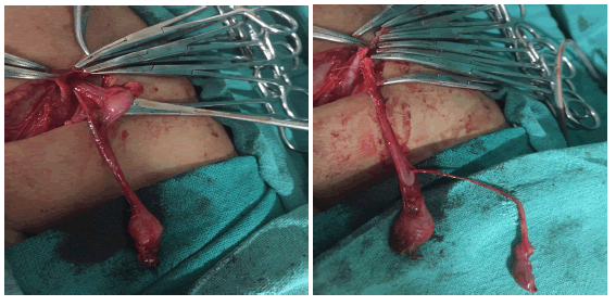
The parents of 2-year-old child came in OPD with a complaint of left UDT. The past medical, family, and psychosocial history was normal. On physical examination, there was a palpable left-sided UDT with a normal right-sided testis. On USG, also triple testis could not be diagnosed, so we thought it to be a usual case of UDT. After routine investigations, the patient was posted for orchidopexy on the left side. During surgery, it was found that there were two testes in the left superficial inguinal pouch. One testis, which had a short vas, was excised, and the other was brought down in the left scrotal sac using the Swanson procedure (Figure 1).
Figure 1. Showing two testes, one with a short vas and the other with a long vas, which underwent orchidopexy on the left side.
Case 2 (2017):
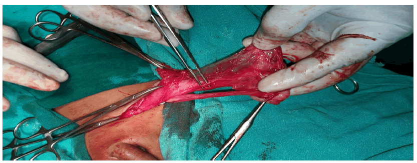
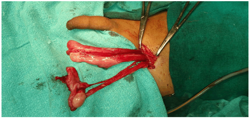
A child of two and a half years reported to the OPD with a complaint of a right-sided undescended testis. On examination left testis was palpable in the scrotal sac, while another swelling was palpable in the inguinal canal (Figures 2 and 3) and on USG, it was suggestive of one more testis, while on the right side testis was absent. The child underwent surgery, and a crossed testicular ectopia was found. The testis in the left inguinal canal was brought down, and orchidopexy was done in the right scrotal sac from the scrotal septum.
Figure 2. Second testis found in sac intraoperatively.
Figure 3. Testis from left inguinal canal with separate Vas with enough length for orchidopexy.
Case 3 (2015):
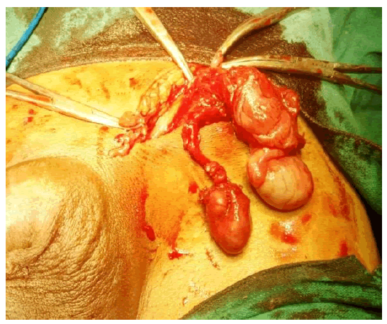
A 47-year-old male patient came to the hospital with a complaint of swelling in the left inguinal region. The patient was diagnosed with a case of left indirect inguinal hernia. On clinical examination of the scrotum, both testes were present, but the left testis was smaller than the right. After investigations, the patient was posted for surgery under spinal anaesthesia. Intra-operatively, it was found that the spermatic cord contained two separate vas deferenses, one was connected to testis in the scrotum and the other testis was present in the inguinal canal (Figure 4). Orchiectomy was performed of the inguinal testes. Hernioplasty was done for inguinal hernia.
Figure 4. Intra-operative view of left inguinal canal showing two testicles.
Case 4 (2008):
A case was reported from our institute, published in the Journal of Pediatric Surgery (JPS) 2008 by Kumar B, Sharma C et al, having left-sided duplex testis and found incidentally during inguinal hernia repair.
Case 5 (2002):
A 4-year-old boy presented with a left undescended testis. On examination left testis was present in the left inguinal region. Exploration revealed two separate testicles within a single tunica vaginalis. The caudal testis was atrophic, but both testis had their own epididymis, vas deferens, and vessels, and the two vas joined together within the operative field to form a common vas. This falls into type 3 of Thum ‘s classification. This case was published in Pediatr Surg Int (2002) 18: 449-450.
Case 6 (1993):
A 2-year-old boy presented with soft swelling in the scrotal and perineal region on the left side. Examination revealed both testis in scrotal sac but on exploration supernumerary testis was seen on left side which was atrophic with complete duplication of the testis, epididymis, and spermatic cord. The excision of atrophic testis along with lymphangiohemangioma in the perineal region. This case was published in Pediatr Surg Int 1994;9:531.
Results and Discussion
Polyorchidism is defined as the presence of more than two testicles. The term "polyorchidism" derives from the Greek root "πoλυ-" (poly-) meaning plenty and the word "Ορχις"(orchis) meaning testicle, and was first used by Lane in 1895.
The first histologically proven case was reported by Ahlfeld in 1880. It is a very rare congenital anomaly believed to result embryologically from an abnormal division of the genital ridge [6]. Embryological theories responsible for polyorchidism include:
- Degeneration of parts of the mesonephric components.
- Duplication of the genital ridge.
- Division of the genital ridge.
There is an increased risk of malignancy if supernumerary testicles are detected [7].
Classification
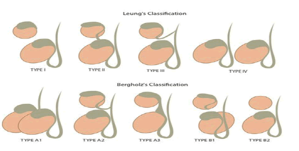
Polyorchidism occurs in two primary forms: Type A and type B (Figure 5).
Figure 5. Types of classification.
Type A: the supernumerary testicle is connected to a vas deferens. These testicles are usually reproductively functional. Type a is further subdivided into:
Type A1: Complete duplication of the testicle, epididymis and vas deferens.
Type A2: The supernumerary testicle has its own epididymis and shares a vas deferens.
Type A3: The supernumerary testicle shares the epididymis and the vas deferens of the other testicles.
Type B: The supernumerary testicle is not connected to a vas deferens and is therefore not reproductively functional. Type b is further subdivided into:
Type B1: The supernumerary testicle has its own epididymis but is not connected to a vas deferens.
Type B2: The supernumerary testicle consists only of testicular tissue.
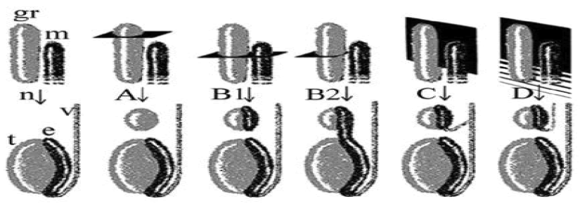
On the basis of the embryologic development, Leung classified polyorchidism into 4 types (Figure 6). In type A, the supernumerary testis lacks an epididymis and vas deferens. It happens when the division separates a small part of the genital ridge not in contact with the mesonephric ducts (rete testis). In type B, the supernumerary testis has its own epididymis. Depending on the degree of division, the supernumerary testis may be connected longitudinally to the epididymis of the normal testis and its vas deferens (B2), or it may lack any connections to the normal testis (B1). The division of the genital ridge occurs in the region where the primordial gonads are attached to the mesonephric ducts (rete testis). In type C, the supernumerary testis has its own epididymis and shares the vas deferens with the regular testis in a parallel fashion. This variant results from incomplete longitudinal division of the genital ridge and the proximal portion of the mesonephric duct. In type D, complete longitudinal duplication of the genital ridge and mesonephric duct occurs, with resultant complete duplication of testes, epididymides, and vas deferens. This type may be associated with an ipsilateral duplicated ureter and is the least common [8,9].
Figure 6. In a normal embryo. Note: (n): At about 6 weeks of embryonic life; (gr): The primordial testis develops from the primitive genital ridge; (m): Medial to the mesonephric duct; At about 8 weeks of embryonic life, the primordial testis; (t): Takes shape; (e): The epididymis; (v): Vas deferens arise from the mesonephric (wolffian) duct.
Differential diagnosis
Possible differential considerations include scrotal hernia, bilobed testicle, crossed testicular ectopia, testicular tumor [10].
Management
The treatment of such patients is still under debate, either removal of supernumerary testis or ectopic testis due to high risk of malignancy (4%-7%) or preservation and long-term follow-up of such patients. Treatment can be based on the presence or absence of vas deferens to supernumerary testicle [11-13].
Conclusion
All the cases were operated in the same institute under the supervision of the same consultants; hence, I took the liberty to compile our center’s cases and add to the literature with our own experience. In our cases, we decided to excise the supernumerary testis as its vas was too short and could not have reached in scrotal sac, and also one testis was brought down by Swanson’s procedure, so it was unnecessary to place two testes in the same dartos pouch. A similar case was reported from our institute in 2008 by Kumar B, et al.
The management protocol which we follow as per our experience is
• Presence or absence of Vas
• Length of Vas
• Location and condition of supernumerary testis in comparison with other two testicles
Declaration of Patient Consent
The authors certify that they have obtained all appropriate patient consent forms. In the form, the legal guardian has given their consent for images and other clinical information to be reported in the journal. The guardian understands that names and initials will not be published and due efforts will be made to conceal patient identity, but anonymity cannot be guaranteed.
Financial Support and Sponsorship
Nil.
Conflicts of Interest
There are no conflicts of interest.
References
- Bergholz R, Wenke K. Polyorchidism: A meta-analysis. J Urol 2009;182(5):2422-2427.
[Crossref] [Google Scholar] [PubMed]
- Rocaâ?Ferrer J, Rodriguez E, Ramírez GA, Moragas C, Sala M. A rare case of polyorchidism in a cat with four intraâ?abdominal testes. Reprod Domest Anim 2015;50(1):172-176.
[Crossref] [Google Scholar] [PubMed]
- Alamsahebpour A, Hidas G, Kaplan A, McAleer IM. Bilateral polyorchidism with diffuse microlithiasis: A case report of an adolescent with 4 testes. Urology 2013;82(6):1421-1423.
[Crossref] [Google Scholar] [PubMed]
- Savas M, Yeni E, Ciftci H, Cece H, Topal U, Utangac MM. Polyorchidism: A threeâ?case report and review of the literature. Andrologia 2010;42(1):57-61.
[Crossref] [Google Scholar] [PubMed]
- Woodward PJ, Schwab CM, Sesterhenn IA. From the archives of the AFIP: Extratesticular scrotal masses: Radiologic pathologic correlation. Radiographics 2003;23(1):215-240.
[Crossref] [Google Scholar] [PubMed]
- Ahlfeld F. Die Missbildungen des Menschen. In: Ahlfeld F, Eds. A Book. Leipzig, Germany: Grunow; 1880:126
- Leun AK. Polyorchidism. Am Fam Phy 1998;38(3):153.
- Amodio JB, Maybody M, Slowotsky C, Fried K, Foresto C. Polyorchidism: Report of 3 cases and review of the literature. J Ultrasound Med 2004;23(7):951-957.
[Crossref] [Google Scholar] [PubMed]
- Barolia DK, Scrhi D. Triple testes-a rare case. Int J Health Sci Res 2015;5(11):403-405.
- Arslanoglu A, Tuncel SA, Hamarat M. Polyorchidism: Color Doppler ultrasonography and magnetic resonance imaging findings. Clin Imaging 2013;37(1):189-191.
[Crossref] [Google Scholar] [PubMed]
- Kumar B, Sharma C, Sinha DD. Supernumerary testis: A case report and review of literature. J Pediat Surg 2008;43(6):9-10.
[Crossref] [Google Scholar] [PubMed]
- Mathur P, Prabhu K, Khamesra HL. Polyorchidism revisited. Pediat Surg Int 2002;18.
- Sharma AK, Mathur P. Polyorchidism. Pediatr Surg Int 1994;9(7):531.
Copyright: © 2025 The Authors. This is an open access article under the terms of the Creative Commons Attribution Non Commercial Share Alike 4.0 (https:// creativecommons.org/licenses/by-nc-sa/4.0/). This is an open access article distributed under the terms of the Creative Commons Attribution License, which permits unrestricted use, distribution, and reproduction in any medium, provided the original work is properly cited.


